2WU4 | pdb_00002wu4

CRYSTAL STRUCTURE OF MOUSE ACETYLCHOLINESTERASE IN COMPLEX WITH FENAMIPHOS AND ORTHO-7


Experimental Data Snapshot

  • Method: X-RAY DIFFRACTION
  • Resolution: 2.40 Å
  • R-Value Free: 
    0.223 (Depositor), 0.208 (DCC) 
  • R-Value Work: 
    0.186 (Depositor), 0.174 (DCC) 
  • R-Value Observed: 
    0.187 (Depositor) 

Starting Model: experimental
View more details

wwPDB Validation 3D Report Full Report

Validation slider image for 2WU4

Ligand Structure Quality Assessment 


This is version 2.3 of the entry. See complete history

Literature

Crystal Structures of Oxime-Bound Fenamiphos-Acetylcholinesterases: Reactivation Involving Flipping of the His447 Ring to Form a Reactive Glu334-His447-Oxime Triad.

Hornberg, A.Artursson, E.Warme, R.Pang, Y.-P.Ekstrom, F.

(2010) Biochem Pharmacol 79: 507

  • DOI: https://doi.org/10.1016/j.bcp.2009.08.027
  • Primary Citation Related Structures: 
    2WU3, 2WU4

  • PubMed Abstract: 

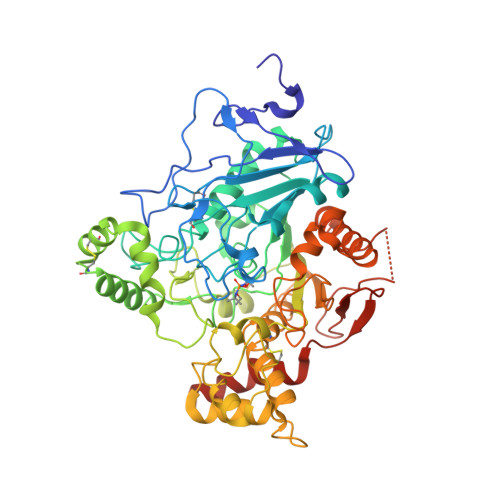
    Organophosphorus insecticides and nerve agents inhibit the vital enzyme acetylcholinesterase by covalently bonding to the catalytic serine residue of the enzyme. Oxime-based reactivators, such as [(E)-[1-[(4-carbamoylpyridin-1-ium-1-yl)methoxymethyl]pyridin-2-ylidene]methyl]-oxoazanium dichloride (HI-6) and 1,7-heptylene-bis-N,N'-2-pyridiniumaldoxime dichloride (Ortho-7), restore the organophosphate-inhibited enzymatic activity by cleaving the phosphorous conjugate. In this article, we report the intermolecular interactions between Mus musculus acetylcholinesterase inhibited by the insecticide fenamiphos (fep-mAChE) and HI-6 or Ortho-7 revealed by a combination of crystallography and kinetics. The crystal structures of the two oxime-bound fep-mAChE complexes show that both oximes interact with the peripheral anionic site involving different conformations of Trp286 and different peripheral-site residues (Tyr124 for HI-6 and Tyr72 for Ortho-7). Moreover, residues at catalytic site of the HI-6-bound fep-mAChE complex adopt conformations that are similar to those in the apo mAChE, whereas significant conformational changes are observed for the corresponding residues in the Ortho-7-bound fep-mAChE complex. Interestingly, flipping of the His447 imidazole ring allows the formation of a hydrogen bonding network among the Glu334-His447-Ortho-7 triad, which presumably deprotonates the Ortho-7 oxime hydroxyl group, increases the nucleophilicity of the oxime group, and leads to cleavage of the phosphorous conjugate. These results offer insights into a detailed reactivation mechanism for the oximes and development of improved reactivators.


  • Organizational Affiliation
    • Swedish Defence Research Agency, Division of CBRN Defence and Security, S-901 82 Umeå, Sweden.

Macromolecule Content 

  • Total Structure Weight: 121.95 kDa 
  • Atom Count: 8,700 
  • Modeled Residue Count: 1,054 
  • Deposited Residue Count: 1,096 
  • Unique protein chains: 1

Macromolecules

Find similar proteins by:|  3D Structure
Entity ID: 1
MoleculeChains  Sequence LengthOrganismDetailsImage
ACETYLCHOLINESTERASE
A, B
548Mus musculusMutation(s): 0 
EC: 3.1.1.7
UniProt & NIH Common Fund Data Resources
Find proteins for P21836 (Mus musculus)
Explore P21836 
Go to UniProtKB:  P21836
IMPC:  MGI:87876
Entity Groups
Sequence Clusters30% Identity50% Identity70% Identity90% Identity95% Identity100% Identity
UniProt GroupP21836
Glycosylation
Glycosylation Sites: 1Go to GlyGen: P21836-1
Sequence Annotations
Expand
Reference Sequence

Small Molecules

Modified Residues  1 Unique
IDChains TypeFormula2D DiagramParent
SXE
Query on SXE
A, B
L-PEPTIDE LINKINGC8 H19 N2 O5 PSER

Experimental Data & Validation

Experimental Data

  • Method: X-RAY DIFFRACTION
  • Resolution: 2.40 Å
  • R-Value Free:  0.223 (Depositor), 0.208 (DCC) 
  • R-Value Work:  0.186 (Depositor), 0.174 (DCC) 
  • R-Value Observed: 0.187 (Depositor) 
Space Group: P 21 21 21
Unit Cell:
Length ( Å )Angle ( ˚ )
a = 79.74α = 90
b = 112.7β = 90
c = 226.69γ = 90
Software Package:
Software NamePurpose
PHENIXrefinement
XDSdata reduction
SCALAdata scaling
CCP4phasing

Structure Validation

View Full Validation Report


Ligand Structure Quality Assessment 


Entry History 

Deposition Data

Revision History  (Full details and data files)

  • Version 1.0: 2009-10-20
    Type: Initial release
  • Version 1.1: 2011-05-08
    Changes: Version format compliance
  • Version 1.2: 2011-07-13
    Changes: Version format compliance
  • Version 1.3: 2018-01-17
    Changes: Data collection
  • Version 2.0: 2019-05-15
    Changes: Data collection, Derived calculations, Polymer sequence
  • Version 2.1: 2020-07-29
    Type: Remediation
    Reason: Carbohydrate remediation
    Changes: Data collection, Derived calculations, Other, Structure summary
  • Version 2.2: 2021-05-12
    Changes: Derived calculations, Structure summary
  • Version 2.3: 2023-12-20
    Changes: Data collection, Database references, Refinement description