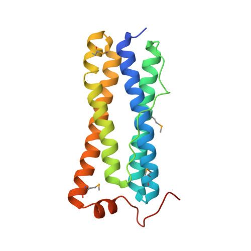
2WTL

Crystal structure of BfrA from M. tuberculosis


Experimental Data Snapshot

  • Method: X-RAY DIFFRACTION
  • Resolution: 2.59 Å
  • R-Value Free: 0.228 
  • R-Value Work: 0.183 
  • R-Value Observed: 0.185 

Starting Model: experimental
View more details

wwPDB Validation   3D Report Full Report


This is version 1.5 of the entry. See complete history


Literature

Crystal Structure of Bfra from Mycobacterium Tuberculosis:Incorporation of Selenomethionine Results in Cleavage and Demetallation of Haem

Gupta, V.Gupta, R.K.Khare, G.Salunke, D.M.Tyagi, A.K.

(2009) PLoS One 4: E8028

  • DOI: https://doi.org/10.1371/journal.pone.0008028
  • Primary Citation of Related Structures:  
    2WTL

  • PubMed Abstract: 

    Emergence of tuberculosis as a global health threat has necessitated an urgent search for new antitubercular drugs entailing determination of 3-dimensional structures of a large number of mycobacterial proteins for structure-based drug design. The essential requirement of ferritins/bacterioferritins (proteins involved in iron storage and homeostasis) for the survival of several prokaryotic pathogens makes these proteins very attractive targets for structure determination and inhibitor design. Bacterioferritins (Bfrs) differ from ferritins in that they have additional noncovalently bound haem groups. The physiological role of haem in Bfrs is not very clear but studies indicate that the haem group is involved in mediating release of iron from Bfr by facilitating reduction of the iron core. To further enhance our understanding, we have determined the crystal structure of the selenomethionyl analog of bacterioferritin A (SeMet-BfrA) from Mycobacterium tuberculosis (Mtb). Unexpectedly, electron density observed in the crystals of SeMet-BfrA analogous to haem location in bacterioferritins, shows a demetallated and degraded product of haem. This unanticipated observation is a consequence of the altered spatial electronic environment around the axial ligands of haem (in lieu of Met52 modification to SeMet52). Furthermore, the structure of Mtb SeMet-BfrA displays a possible lost protein interaction with haem propionates due to formation of a salt bridge between Arg53-Glu57, which appears to be unique to Mtb BfrA, resulting in slight modulation of haem binding pocket in this organism. The crystal structure of Mtb SeMet-BfrA provides novel leads to physiological function of haem in Bfrs. If validated as a drug target, it may also serve as a scaffold for designing specific inhibitors. In addition, this study provides evidence against the general belief that a selenium derivative of a protein represents its true physiological native structure.


  • Organizational Affiliation

    Department of Biochemistry, University of Delhi South Campus, New Delhi, India.


Macromolecules
Find similar proteins by:  (by identity cutoff)  |  3D Structure
Entity ID: 1
MoleculeChains Sequence LengthOrganismDetailsImage
BACTERIOFERRITIN
A, B, C, D, E
A, B, C, D, E, F
173Mycobacterium tuberculosis H37RvMutation(s): 0 
EC: 1.16.3.1
UniProt
Find proteins for P9WPQ9 (Mycobacterium tuberculosis (strain ATCC 25618 / H37Rv))
Explore P9WPQ9 
Go to UniProtKB:  P9WPQ9
Entity Groups  
Sequence Clusters30% Identity50% Identity70% Identity90% Identity95% Identity100% Identity
UniProt GroupP9WPQ9
Sequence Annotations
Expand
  • Reference Sequence
Small Molecules
Ligands 3 Unique
IDChains Name / Formula / InChI Key2D Diagram3D Interactions
FE
Query on FE

Download Ideal Coordinates CCD File 
G [auth A]
H [auth A]
L [auth B]
M [auth B]
N [auth C]
G [auth A],
H [auth A],
L [auth B],
M [auth B],
N [auth C],
O [auth C],
Q [auth D],
R [auth D],
S [auth E],
T [auth E],
W [auth F],
X [auth F]
FE (III) ION
Fe
VTLYFUHAOXGGBS-UHFFFAOYSA-N
UNL
Query on UNL

Download Ideal Coordinates CCD File 
K [auth A],
P [auth C],
V [auth E]
Unknown ligand
VTLYFUHAOXGGBS-UHFFFAOYSA-N
UNX
Query on UNX

Download Ideal Coordinates CCD File 
I [auth A],
J [auth A],
U [auth E]
UNKNOWN ATOM OR ION
X
Modified Residues  1 Unique
IDChains TypeFormula2D DiagramParent
MSE
Query on MSE
A, B, C, D, E
A, B, C, D, E, F
L-PEPTIDE LINKINGC5 H11 N O2 SeMET
Experimental Data & Validation

Experimental Data

  • Method: X-RAY DIFFRACTION
  • Resolution: 2.59 Å
  • R-Value Free: 0.228 
  • R-Value Work: 0.183 
  • R-Value Observed: 0.185 
  • Space Group: I 4
Unit Cell:
Length ( Å )Angle ( ˚ )
a = 125.96α = 90
b = 125.96β = 90
c = 175.84γ = 90
Software Package:
Software NamePurpose
PHENIXrefinement
AUTOMARdata reduction
AUTOMARdata scaling
PHENIXphasing

Structure Validation

View Full Validation Report


Entry History 

Deposition Data

Revision History  (Full details and data files)

  • Version 1.0: 2009-12-15
    Type: Initial release
  • Version 1.1: 2011-05-08
    Changes: Version format compliance
  • Version 1.2: 2011-07-13
    Changes: Version format compliance
  • Version 1.3: 2019-07-24
    Changes: Data collection, Derived calculations
  • Version 1.4: 2023-12-20
    Changes: Data collection, Database references, Derived calculations, Other, Refinement description
  • Version 1.5: 2024-10-23
    Changes: Structure summary