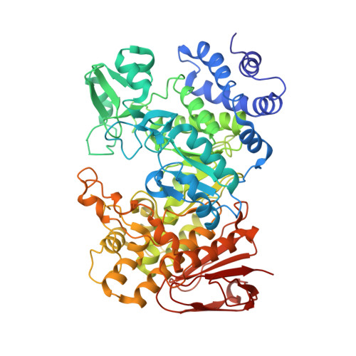
The Apo Structure of Sucrose Hydrolase from Xanthomonas Campestris Pv. Campestris Shows an Open Active-Site Groove
Champion, E., Remaud-Simeon, M., Skov, L.K., Kastrup, J.S., Gajhede, M., Mirza, O.(2009) Acta Crystallogr D Biol Crystallogr 65: 1309
- PubMed: 19966417
- DOI: https://doi.org/10.1107/S0907444909040311
- Primary Citation of Related Structures:
2WPG - PubMed Abstract:
Glycoside hydrolase family 13 (GH-13) mainly contains starch-degrading or starch-modifying enzymes. Sucrose hydrolases utilize sucrose instead of amylose as the primary glucosyl donor. Here, the catalytic properties and X-ray structure of sucrose hydrolase from Xanthomonas campestris pv. campestris are reported. Sucrose hydrolysis catalyzed by the enzyme follows Michaelis-Menten kinetics, with a K(m) of 60.7 mM and a k(cat) of 21.7 s(-1). The structure of the enzyme was solved at a resolution of 1.9 A in the resting state with an empty active site. This represents the first apo structure from subfamily 4 of GH-13. Comparisons with structures of the highly similar sucrose hydrolase from X. axonopodis pv. glycines most notably showed that residues Arg516 and Asp138, which form a salt bridge in the X. axonopodis sucrose complex and define part of the subsite -1 glucosyl-binding determinants, are not engaged in salt-bridge formation in the resting X. campestris enzyme. In the absence of the salt bridge an opening is created which gives access to subsite -1 from the ;nonreducing' end. Binding of the glucosyl moiety in subsite -1 is therefore likely to induce changes in the conformation of the active-site cleft of the X. campestris enzyme. These changes lead to salt-bridge formation that shortens the groove. Additionally, this finding has implications for understanding the molecular mechanism of the closely related subfamily 4 glucosyl transferase amylosucrase, as it indicates that sucrose could enter the active site from the ;nonreducing' end during the glucan-elongation cycle.
- Laboratoire d'Ingénierie des Systèmes Biologiques et des Procédés, Toulouse, France.
Organizational Affiliation:


