2WNG | pdb_00002wng

complete extracellular structure of human signal regulatory protein (SIRP) alpha


Experimental Data Snapshot

  • Method: X-RAY DIFFRACTION
  • Resolution: 2.49 Å
  • R-Value Free: 
    0.271 (Depositor), 0.290 (DCC) 
  • R-Value Work: 
    0.219 (Depositor), 0.220 (DCC) 
  • R-Value Observed: 
    0.221 (Depositor) 

Starting Models: experimental
View more details

wwPDB Validation   3D Report Full Report


Ligand Structure Quality Assessment 

Created with Raphaël 2.3.0Worse 01 BetterLigand structure goodness of fit to experimental dataBest fitted NAGClick on this verticalbar to view details

This is version 1.7 of the entry. See complete history


Literature

Structure of Signal-Regulatory Protein Alpha: A Link to Antigen Receptor Evolution.

Hatherley, D.Graham, S.C.Harlos, K.Stuart, D.I.Barclay, A.N.

(2009) J Biological Chem 284: 26613

  • DOI: https://doi.org/10.1074/jbc.M109.017566
  • Primary Citation of Related Structures:  
    2WNG

  • PubMed Abstract: 

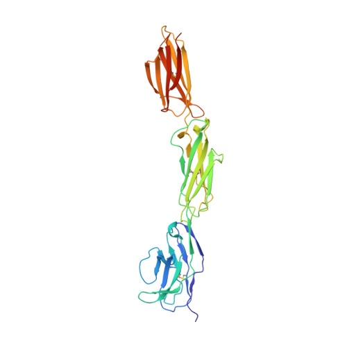
    Signal-regulatory protein alpha (SIRPalpha) is a myeloid membrane receptor that interacts with the membrane protein CD47, a marker of self. We have solved the structure of the complete extracellular portion of SIRPalpha, comprising three immunoglobulin superfamily domains, by x-ray crystallography to 2.5 A resolution. These data, together with previous data on the N-terminal domain and its ligand CD47 (possessing a single immunoglobulin superfamily domain), show that the CD47-SIRPalpha interaction will span a distance of around 14 nm between interacting cells, comparable with that of an immunological synapse. The N-terminal (V-set) domain mediates binding to CD47, and the two others are found to be constant (C1-set) domains. C1-set domains are restricted to proteins involved in vertebrate antigen recognition: T cell antigen receptors, immunoglobulins, major histocompatibility complex antigens, tapasin, and beta2-microglobulin. The domains of SIRPalpha (domains 2 and 3) are structurally more similar to C1-set domains than any cell surface protein not involved in antigen recognition. This strengthens the suggestion from sequence analysis that SIRP is evolutionarily closely related to antigen recognition proteins.


  • Organizational Affiliation

    Sir William Dunn School of Pathology, University of Oxford, Oxford OX1 3RE, United Kingdom.


Macromolecules
Find similar proteins by:  (by identity cutoff)  |  3D Structure
Entity ID: 1
MoleculeChains Sequence LengthOrganismDetailsImage
TYROSINE-PROTEIN PHOSPHATASE NON-RECEPTOR TYPE SUBSTRATE 1327Homo sapiensMutation(s): 0 
UniProt & NIH Common Fund Data Resources
Find proteins for P78324 (Homo sapiens)
Explore P78324 
Go to UniProtKB:  P78324
PHAROS:  P78324
GTEx:  ENSG00000198053 
Entity Groups  
Sequence Clusters30% Identity50% Identity70% Identity90% Identity95% Identity100% Identity
UniProt GroupP78324
Glycosylation
Glycosylation Sites: 1Go to GlyGen: P78324-1
Sequence Annotations
Expand
  • Reference Sequence
Small Molecules
Ligands 1 Unique
IDChains Name / Formula / InChI Key2D Diagram3D Interactions
NAG
Query on NAG

Download Ideal Coordinates CCD File 
B [auth A]2-acetamido-2-deoxy-beta-D-glucopyranose
C8 H15 N O6
OVRNDRQMDRJTHS-FMDGEEDCSA-N
Experimental Data & Validation

Experimental Data

  • Method: X-RAY DIFFRACTION
  • Resolution: 2.49 Å
  • R-Value Free:  0.271 (Depositor), 0.290 (DCC) 
  • R-Value Work:  0.219 (Depositor), 0.220 (DCC) 
  • R-Value Observed: 0.221 (Depositor) 
Space Group: P 21 21 21
Unit Cell:
Length ( Å )Angle ( ˚ )
a = 47.41α = 90
b = 84.37β = 90
c = 98.86γ = 90
Software Package:
Software NamePurpose
BUSTER-TNTrefinement
XDSdata reduction
XSCALEdata scaling
MOLREPphasing

Structure Validation

View Full Validation Report


Ligand Structure Quality Assessment 

Created with Raphaël 2.3.0Worse 01 BetterLigand structure goodness of fit to experimental dataBest fitted NAGClick on this verticalbar to view details

Entry History 

Deposition Data

Revision History  (Full details and data files)

  • Version 1.0: 2009-07-21
    Type: Initial release
  • Version 1.1: 2011-05-08
    Changes: Version format compliance
  • Version 1.2: 2011-07-13
    Changes: Version format compliance
  • Version 1.3: 2019-04-03
    Changes: Data collection, Derived calculations, Other, Source and taxonomy
  • Version 1.4: 2019-05-08
    Changes: Data collection, Experimental preparation
  • Version 1.5: 2020-07-29
    Type: Remediation
    Reason: Carbohydrate remediation
    Changes: Data collection, Derived calculations, Other, Structure summary
  • Version 1.6: 2023-12-13
    Changes: Data collection, Database references, Refinement description, Structure summary
  • Version 1.7: 2024-11-06
    Changes: Structure summary