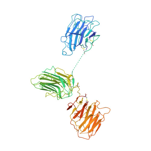
2WJS

Crystal structure of the LG1-3 region of the laminin alpha2 chain


Experimental Data Snapshot

  • Method: X-RAY DIFFRACTION
  • Resolution: 2.80 Å
  • R-Value Free: 0.266 
  • R-Value Work: 0.212 
  • R-Value Observed: 0.212 

Starting Model: experimental
View more details

wwPDB Validation   3D Report Full Report


Ligand Structure Quality Assessment 


This is version 1.4 of the entry. See complete history


Literature

Crystal Structure of the Lg1-3 Region of the Laminin {Alpha}2 Chain.

Carafoli, F.Clout, N.J.Hohenester, E.

(2009) J Biol Chem 284: 22786

  • DOI: https://doi.org/10.1074/jbc.M109.026658
  • Primary Citation of Related Structures:  
    2WJS

  • PubMed Abstract: 

    Laminins are large heterotrimeric glycoproteins with many essential functions in basement membrane assembly and function. Cell adhesion to laminins is mediated by a tandem of five laminin G-like (LG) domains at the C terminus of the alpha chain. Integrin binding requires an intact LG1-3 region, as well as contributions from the coiled coil formed by the alpha, beta, and gamma chains. We have determined the crystal structure at 2.8-A resolution of the LG1-3 region of the laminin alpha2 chain (alpha 2LG1-3). The three LG domains adopt typical beta-sandwich folds, with canonical calcium binding sites in LG1 and LG2. LG2 and LG3 interact through a substantial interface, but LG1 is completely dissociated from the LG2-3 pair. We suggest that the missing gamma chain tail may be required to stabilize the interaction between LG1 and LG2-3 in the biologically active conformation. A global analysis of N-linked glycosylation sites shows that the beta-sandwich faces of LG1 are free of carbohydrate modifications in all five laminin alpha chains, suggesting that these surfaces may harbor the integrin binding site. The alpha 2LG1-3 structure provides the first atomic view of the integrin binding region of laminins.


  • Organizational Affiliation

    Department of Life Sciences, Imperial College London, London SW7 2AZ, United Kingdom.


Macromolecules
Find similar proteins by:  (by identity cutoff)  |  3D Structure
Entity ID: 1
MoleculeChains Sequence LengthOrganismDetailsImage
LAMININ SUBUNIT ALPHA-2608Mus musculusMutation(s): 0 
UniProt
Find proteins for Q60675 (Mus musculus)
Explore Q60675 
Go to UniProtKB:  Q60675
Entity Groups  
Sequence Clusters30% Identity50% Identity70% Identity90% Identity95% Identity100% Identity
UniProt GroupQ60675
Glycosylation
Glycosylation Sites: 4Go to GlyGen: Q60675-1
Sequence Annotations
Expand
  • Reference Sequence
Experimental Data & Validation

Experimental Data

  • Method: X-RAY DIFFRACTION
  • Resolution: 2.80 Å
  • R-Value Free: 0.266 
  • R-Value Work: 0.212 
  • R-Value Observed: 0.212 
  • Space Group: P 65
Unit Cell:
Length ( Å )Angle ( ˚ )
a = 138.829α = 90
b = 138.829β = 90
c = 73.895γ = 120
Software Package:
Software NamePurpose
CNSrefinement
MOSFLMdata reduction
SCALAdata scaling
PHASERphasing

Structure Validation

View Full Validation Report


Ligand Structure Quality Assessment 


Entry History 

Deposition Data

Revision History  (Full details and data files)

  • Version 1.0: 2009-06-23
    Type: Initial release
  • Version 1.1: 2011-05-08
    Changes: Version format compliance
  • Version 1.2: 2011-07-13
    Changes: Version format compliance
  • Version 1.3: 2020-07-29
    Type: Remediation
    Reason: Carbohydrate remediation
    Changes: Data collection, Derived calculations, Other, Structure summary
  • Version 1.4: 2023-12-13
    Changes: Data collection, Database references, Refinement description, Structure summary