2WD7 | pdb_00002wd7

PTERIDINE REDUCTASE 1 (PTR1) FROM TRYPANOSOMA BRUCEI IN COMPLEX WITH NADP AND DDD00066750


Experimental Data Snapshot

  • Method: X-RAY DIFFRACTION
  • Resolution: 1.90 Å
  • R-Value Free: 
    0.202 (Depositor), 0.201 (DCC) 
  • R-Value Work: 
    0.155 (Depositor), 0.154 (DCC) 
  • R-Value Observed: 
    0.157 (Depositor) 

Starting Model: experimental
View more details

wwPDB Validation 3D Report Full Report

Validation slider image for 2WD7

Ligand Structure Quality Assessment 


This is version 1.3 of the entry. See complete history

Literature

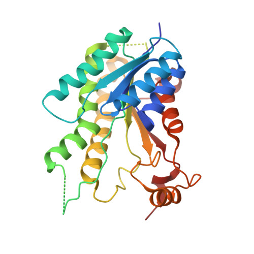
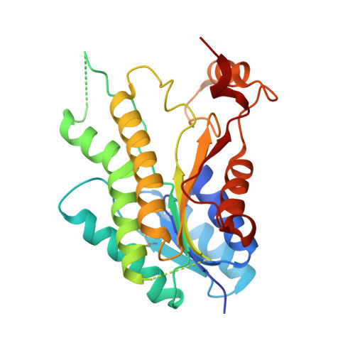
One Scaffold, Three Binding Modes: Novel and Selective Pteridine Reductase 1 Inhibitors Derived from Fragment Hits Discovered by Virtual Screening.

Mpamhanga, C.P.Spinks, D.Tulloch, L.B.Shanks, E.J.Robinson, D.A.Collie, I.T.Fairlamb, A.H.Wyatt, P.G.Frearson, J.A.Hunter, W.N.Gilbert, I.H.Brenk, R.

(2009) J Med Chem 52: 4454

  • DOI: https://doi.org/10.1021/jm900414x
  • Primary Citation Related Structures: 
    2WD7, 2WD8, 3GN1, 3GN2

  • PubMed Abstract: 

    The enzyme pteridine reductase 1 (PTR1) is a potential target for new compounds to treat human African trypanosomiasis. A virtual screening campaign for fragments inhibiting PTR1 was carried out. Two novel chemical series were identified containing aminobenzothiazole and aminobenzimidazole scaffolds, respectively. One of the hits (2-amino-6-chloro-benzimidazole) was subjected to crystal structure analysis and a high resolution crystal structure in complex with PTR1 was obtained, confirming the predicted binding mode. However, the crystal structures of two analogues (2-amino-benzimidazole and 1-(3,4-dichloro-benzyl)-2-amino-benzimidazole) in complex with PTR1 revealed two alternative binding modes. In these complexes, previously unobserved protein movements and water-mediated protein-ligand contacts occurred, which prohibited a correct prediction of the binding modes. On the basis of the alternative binding mode of 1-(3,4-dichloro-benzyl)-2-amino-benzimidazole, derivatives were designed and selective PTR1 inhibitors with low nanomolar potency and favorable physicochemical properties were obtained.


  • Organizational Affiliation
    • Division of Biological Chemistry and Drug Discovery, College of Life Sciences, University of Dundee, Dundee DD1 5EH, UK.

Macromolecule Content 

  • Total Structure Weight: 117.69 kDa 
  • Atom Count: 8,511 
  • Modeled Residue Count: 995 
  • Deposited Residue Count: 1,072 
  • Unique protein chains: 2

Macromolecules

Find similar proteins by:|  3D Structure
Entity ID: 1
MoleculeChains  Sequence LengthOrganismDetailsImage
PTERIDINE REDUCTASE268Trypanosoma brucei bruceiMutation(s): 0 
EC: 1.5.1.33
UniProt
Find proteins for O76290 (Trypanosoma brucei brucei)
Explore O76290 
Go to UniProtKB:  O76290
Entity Groups
Sequence Clusters30% Identity50% Identity70% Identity90% Identity95% Identity100% Identity
UniProt GroupO76290
Sequence Annotations
Expand
Reference Sequence
Find similar proteins by:|  3D Structure
Entity ID: 2
MoleculeChains  Sequence LengthOrganismDetailsImage
PTERIDINE REDUCTASE
B, C, D
268Trypanosoma brucei bruceiMutation(s): 0 
EC: 1.5.1.33
UniProt
Find proteins for O76290 (Trypanosoma brucei brucei)
Explore O76290 
Go to UniProtKB:  O76290
Entity Groups
Sequence Clusters30% Identity50% Identity70% Identity90% Identity95% Identity100% Identity
UniProt GroupO76290
Sequence Annotations
Expand
Reference Sequence

Small Molecules

Ligands 2 Unique
IDChains Name / Formula / InChI Key2D Diagram3D Interactions
NAP

Query on NAP


Download:Ideal Coordinates CCD File
E [auth A],
G [auth B],
I [auth C],
K [auth D]
NADP NICOTINAMIDE-ADENINE-DINUCLEOTIDE PHOSPHATE
C21 H28 N7 O17 P3
XJLXINKUBYWONI-NNYOXOHSSA-N
VGD

Query on VGD


Download:Ideal Coordinates CCD File
F [auth A],
H [auth B],
J [auth C],
L [auth D]
6-chloro-1H-benzimidazol-2-amine
C7 H6 Cl N3
PDOCNPCPPLPXRV-UHFFFAOYSA-N
Modified Residues  1 Unique
IDChains TypeFormula2D DiagramParent
CSX
Query on CSX
B, C, D
L-PEPTIDE LINKINGC3 H7 N O3 SCYS

Experimental Data & Validation

Experimental Data

  • Method: X-RAY DIFFRACTION
  • Resolution: 1.90 Å
  • R-Value Free:  0.202 (Depositor), 0.201 (DCC) 
  • R-Value Work:  0.155 (Depositor), 0.154 (DCC) 
  • R-Value Observed: 0.157 (Depositor) 
Space Group: P 1 21 1
Unit Cell:
Length ( Å )Angle ( ˚ )
a = 74.677α = 90
b = 89.89β = 115.48
c = 82.695γ = 90
Software Package:
Software NamePurpose
REFMACrefinement
MOSFLMdata reduction
SCALAdata scaling
MOLREPphasing

Structure Validation

View Full Validation Report


Ligand Structure Quality Assessment 


Entry History 

Deposition Data

Revision History  (Full details and data files)

  • Version 1.0: 2009-06-30
    Type: Initial release
  • Version 1.1: 2011-07-13
    Changes: Advisory, Version format compliance
  • Version 1.2: 2012-10-10
    Changes: Database references, Derived calculations, Non-polymer description
  • Version 1.3: 2023-12-13
    Changes: Data collection, Database references, Derived calculations, Other, Refinement description