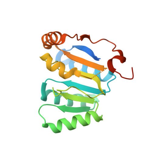
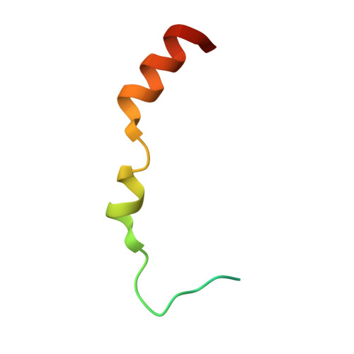
2WAX

Structure of the human DDX6 C-terminal domain in complex with an EDC3- FDF peptide


Experimental Data Snapshot

  • Method: X-RAY DIFFRACTION
  • Resolution: 2.30 Å
  • R-Value Free: 0.260 
  • R-Value Work: 0.190 
  • R-Value Observed: 0.193 

Starting Model: experimental
View more details

wwPDB Validation   3D Report Full Report


Ligand Structure Quality Assessment 


This is version 1.3 of the entry. See complete history

Re-refinement Note

A newer entry is available that reflects an alternative modeling of the original data: 6S8S


Literature

Structural Basis for the Mutually Exclusive Anchoring of P Body Components Edc3 and Tral to the Dead Box Protein Ddx6/Me31B.

Tritschler, F.Braun, J.E.Eulalio, A.Truffault, V.Izaurralde, E.Weichenrieder, O.

(2009) Mol Cell 33: 661

  • DOI: https://doi.org/10.1016/j.molcel.2009.02.014
  • Primary Citation of Related Structures:  
    2WAX, 2WAY

  • PubMed Abstract: 

    The DEAD box helicase DDX6/Me31B functions in translational repression and mRNA decapping. How particular RNA helicases are recruited specifically to distinct functional complexes is poorly understood. We present the crystal structure of the DDX6 C-terminal RecA-like domain bound to a highly conserved FDF sequence motif in the decapping activator EDC3. The FDF peptide adopts an alpha-helical conformation upon binding to DDX6, occupying a shallow groove opposite to the DDX6 surface involved in RNA binding and ATP hydrolysis. Mutagenesis of Me31B shows the relevance of the FDF interaction surface both for Me31B's accumulation in P bodies and for its ability to repress the expression of bound mRNAs. The translational repressor Tral contains a similar FDF motif. Together with mutational and competition studies, the structure reveals why the interactions of Me31B with EDC3 and Tral are mutually exclusive and how the respective decapping and translational repressor complexes might hook onto an mRNA substrate.


  • Organizational Affiliation

    Max Planck Institute for Developmental Biology, Tübingen, Germany.


Macromolecules
Find similar proteins by:  (by identity cutoff)  |  3D Structure
Entity ID: 1
MoleculeChains Sequence LengthOrganismDetailsImage
ATP-DEPENDENT RNA HELICASE DDX6
A, C
193Homo sapiensMutation(s): 0 
EC: 3.6.1 (PDB Primary Data), 3.6.4.13 (UniProt)
UniProt & NIH Common Fund Data Resources
Find proteins for P26196 (Homo sapiens)
Explore P26196 
Go to UniProtKB:  P26196
PHAROS:  P26196
GTEx:  ENSG00000110367 
Entity Groups  
Sequence Clusters30% Identity50% Identity70% Identity90% Identity95% Identity100% Identity
UniProt GroupP26196
Sequence Annotations
Expand
  • Reference Sequence
Find similar proteins by:  (by identity cutoff)  |  3D Structure
Entity ID: 2
MoleculeChains Sequence LengthOrganismDetailsImage
ENHANCER OF MRNA-DECAPPING PROTEIN 3
B, D
44Homo sapiensMutation(s): 0 
UniProt & NIH Common Fund Data Resources
Find proteins for Q96F86 (Homo sapiens)
Explore Q96F86 
Go to UniProtKB:  Q96F86
PHAROS:  Q96F86
GTEx:  ENSG00000179151 
Entity Groups  
Sequence Clusters30% Identity50% Identity70% Identity90% Identity95% Identity100% Identity
UniProt GroupQ96F86
Sequence Annotations
Expand
  • Reference Sequence
Small Molecules
Ligands 2 Unique
IDChains Name / Formula / InChI Key2D Diagram3D Interactions
CXS
Query on CXS

Download Ideal Coordinates CCD File 
E [auth A]
F [auth A]
G [auth A]
K [auth C]
L [auth C]
E [auth A],
F [auth A],
G [auth A],
K [auth C],
L [auth C],
N [auth D]
3-CYCLOHEXYL-1-PROPYLSULFONIC ACID
C9 H19 N O3 S
PJWWRFATQTVXHA-UHFFFAOYSA-N
PO4
Query on PO4

Download Ideal Coordinates CCD File 
H [auth A],
I [auth A],
J [auth B],
M [auth C]
PHOSPHATE ION
O4 P
NBIIXXVUZAFLBC-UHFFFAOYSA-K
Experimental Data & Validation

Experimental Data

  • Method: X-RAY DIFFRACTION
  • Resolution: 2.30 Å
  • R-Value Free: 0.260 
  • R-Value Work: 0.190 
  • R-Value Observed: 0.193 
  • Space Group: C 1 2 1
Unit Cell:
Length ( Å )Angle ( ˚ )
a = 172.63α = 90
b = 47.85β = 96.35
c = 65.78γ = 90
Software Package:
Software NamePurpose
REFMACrefinement
XDSdata reduction
XSCALEdata scaling
PHASERphasing

Structure Validation

View Full Validation Report


Ligand Structure Quality Assessment 


Entry History 

Deposition Data

Revision History  (Full details and data files)

  • Version 1.0: 2009-03-24
    Type: Initial release
  • Version 1.1: 2011-05-08
    Changes: Version format compliance
  • Version 1.2: 2011-07-13
    Changes: Version format compliance
  • Version 1.3: 2023-12-13
    Changes: Data collection, Database references, Derived calculations, Other, Refinement description