2W8D | pdb_00002w8d

Distinct and essential morphogenic functions for wall- and lipo- teichoic acids in Bacillus subtilis


Experimental Data Snapshot

  • Method: X-RAY DIFFRACTION
  • Resolution: 2.35 Å
  • R-Value Free: 
    0.235 (Depositor), 0.257 (DCC) 
  • R-Value Work: 
    0.170 (Depositor), 0.196 (DCC) 
  • R-Value Observed: 
    0.174 (Depositor) 

wwPDB Validation 3D Report Full Report

Validation slider image for 2W8D

This is version 1.2 of the entry. See complete history

Literature

Distinct and Essential Morphogenic Functions for Wall- and Lipo-Teichoic Acids in Bacillus Subtilis

Schirner, K.Marles-Wright, J.Lewis, R.J.Errington, J.

(2009) EMBO J 28: 830

  • DOI: https://doi.org/10.1038/emboj.2009.25
  • Primary Citation Related Structures: 
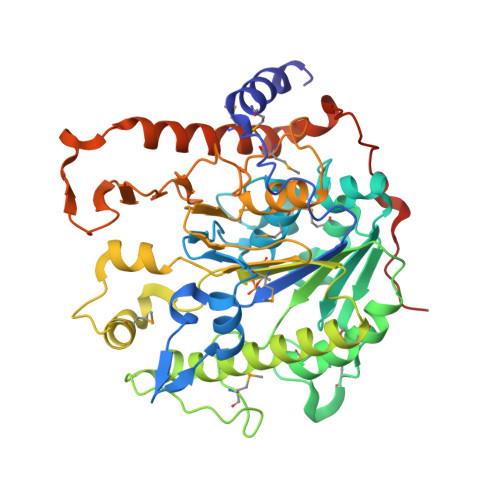
    2W8D

  • PubMed Abstract: 

    Teichoic acids (TAs) are anionic polymers that constitute a major component of the cell wall in most Gram-positive bacteria. Despite decades of study, their function has remained unclear. TAs are covalently linked either to the cell wall peptidoglycan (wall TA (WTA)) or to the membrane (lipo-TA (LTA)). We have characterized the key enzyme of LTA synthesis in Bacillus subtilis, LTA synthase (LtaS). We show that LTA is needed for divalent cation homoeostasis and that its absence has severe effects on cell morphogenesis and cell division. Inactivation of both LTA and WTA is lethal and comparison of the individual mutants suggests that they have differentiated roles in elongation (WTA) and division (LTA). B. subtilis has four ltaS paralogues and we show how their roles are partially differentiated. Two paralogues have a redundant role in LTA synthesis during sporulation and their absence gives a novel absolute block in sporulation. The crystal structure of the extracytoplasmic part of LtaS, solved at 2.4-A resolution, reveals a phosphorylated threonine residue, which provides clues about the catalytic mechanism and identifies the active site of the enzyme.


  • Organizational Affiliation
    • Centre for Bacterial Cell Biology, Institute for Cell and Molecular Biosciences, The Medical School, Newcastle University, Newcastle upon Tyne, UK.

Macromolecule Content 

  • Total Structure Weight: 101.46 kDa 
  • Atom Count: 7,060 
  • Modeled Residue Count: 841 
  • Deposited Residue Count: 872 
  • Unique protein chains: 1

Macromolecules

Find similar proteins by:|  3D Structure
Entity ID: 1
MoleculeChains  Sequence LengthOrganismDetailsImage
PROCESSED GLYCEROL PHOSPHATE LIPOTEICHOIC ACID SYNTHASE 2
A, B
436Bacillus subtilisMutation(s): 0 
EC: 2.7.8
Membrane Entity: Yes 
UniProt
Find proteins for O34952 (Bacillus subtilis (strain 168))
Explore O34952 
Go to UniProtKB:  O34952
Entity Groups
Sequence Clusters30% Identity50% Identity70% Identity90% Identity95% Identity100% Identity
UniProt GroupO34952
Sequence Annotations
Expand
Reference Sequence

Experimental Data & Validation

Experimental Data

  • Method: X-RAY DIFFRACTION
  • Resolution: 2.35 Å
  • R-Value Free:  0.235 (Depositor), 0.257 (DCC) 
  • R-Value Work:  0.170 (Depositor), 0.196 (DCC) 
  • R-Value Observed: 0.174 (Depositor) 
Space Group: P 1 21 1
Unit Cell:
Length ( Å )Angle ( ˚ )
a = 56.627α = 90
b = 54.408β = 90.97
c = 140.783γ = 90
Software Package:
Software NamePurpose
REFMACrefinement
MOSFLMdata reduction
SCALAdata scaling
SHELXphasing

Structure Validation

View Full Validation Report


Entry History 

Deposition Data

Revision History  (Full details and data files)

  • Version 1.0: 2009-03-03
    Type: Initial release
  • Version 1.1: 2011-07-13
    Changes: Advisory, Refinement description, Version format compliance
  • Version 1.2: 2024-10-23
    Changes: Data collection, Database references, Derived calculations, Other, Structure summary