2W83

Crystal structure of the ARF6 GTPase in complex with a specific effector, JIP4


Experimental Data Snapshot

  • Method: X-RAY DIFFRACTION
  • Resolution: 1.93 Å
  • R-Value Free: 0.238 
  • R-Value Work: 0.214 
  • R-Value Observed: 0.217 

Starting Model: experimental
View more details

wwPDB Validation   3D Report Full Report


Ligand Structure Quality Assessment 


This is version 1.3 of the entry. See complete history


Literature

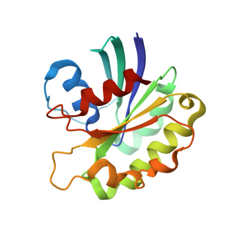
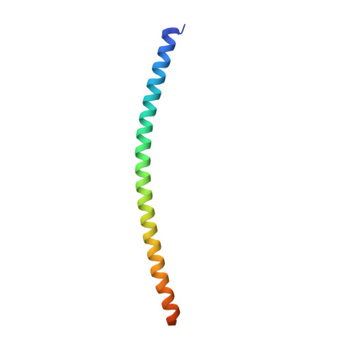
The Structural Basis of Arf Effector Specificity: The Crystal Structure of Arf6 in a Complex with Jip4.

Isabet, T.Montagnac, G.Regazzoni, K.Raynal, B.El Khadali, F.England, P.Franco, M.Chavrier, P.Houdusse, A.Menetrey, J.

(2009) EMBO J 28: 2835

  • DOI: https://doi.org/10.1038/emboj.2009.209
  • Primary Citation of Related Structures:  
    2W83

  • PubMed Abstract: 

    The JNK-interacting proteins, JIP3 and JIP4, are specific effectors of the small GTP-binding protein ARF6. The interaction of ARF6-GTP with the second leucine zipper (LZII) domains of JIP3/JIP4 regulates the binding of JIPs to kinesin-1 and dynactin. Here, we report the crystal structure of ARF6-GTP bound to the JIP4-LZII at 1.9 A resolution. The complex is a heterotetramer with dyad symmetry arranged in an ARF6-(JIP4)(2)-ARF6 configuration. Comparison of the ARF6-JIP4 interface with the equivalent region of ARF1 shows the structural basis of JIP4's specificity for ARF6. Using site-directed mutagenesis and surface plasmon resonance, we further show that non-conserved residues at the switch region borders are the key structural determinants of JIP4 specificity. A structure-derived model of the association of the ARF6-JIP3/JIP4 complex with membranes shows that the JIP4-LZII coiled-coil should lie along the membrane to prevent steric hindrances, resulting in only one ARF6 molecule bound. Such a heterotrimeric complex gives insights to better understand the ARF6-mediated motor switch regulatory function.


  • Organizational Affiliation

    Institut Curie, Centre de Recherche, Paris, France.


Macromolecules
Find similar proteins by:  (by identity cutoff)  |  3D Structure
Entity ID: 1
MoleculeChains Sequence LengthOrganismDetailsImage
ADP-RIBOSYLATION FACTOR 6
A, B, E
165Homo sapiensMutation(s): 1 
UniProt & NIH Common Fund Data Resources
Find proteins for P62330 (Homo sapiens)
Explore P62330 
Go to UniProtKB:  P62330
GTEx:  ENSG00000165527 
Entity Groups  
Sequence Clusters30% Identity50% Identity70% Identity90% Identity95% Identity100% Identity
UniProt GroupP62330
Sequence Annotations
Expand
  • Reference Sequence
Find similar proteins by:  (by identity cutoff)  |  3D Structure
Entity ID: 2
MoleculeChains Sequence LengthOrganismDetailsImage
C-JUN-AMINO-TERMINAL KINASE-INTERACTING PROTEIN 4
C, D
77Homo sapiensMutation(s): 0 
UniProt & NIH Common Fund Data Resources
Find proteins for O60271 (Homo sapiens)
Explore O60271 
Go to UniProtKB:  O60271
GTEx:  ENSG00000008294 
Entity Groups  
Sequence Clusters30% Identity50% Identity70% Identity90% Identity95% Identity100% Identity
UniProt GroupO60271
Sequence Annotations
Expand
  • Reference Sequence
Small Molecules
Ligands 4 Unique
IDChains Name / Formula / InChI Key2D Diagram3D Interactions
GTP
Query on GTP

Download Ideal Coordinates CCD File 
F [auth A],
J [auth B],
N [auth E]
GUANOSINE-5'-TRIPHOSPHATE
C10 H16 N5 O14 P3
XKMLYUALXHKNFT-UUOKFMHZSA-N
GOL
Query on GOL

Download Ideal Coordinates CCD File 
I [auth A]GLYCEROL
C3 H8 O3
PEDCQBHIVMGVHV-UHFFFAOYSA-N
DIO
Query on DIO

Download Ideal Coordinates CCD File 
H [auth A]
L [auth C]
M [auth C]
P [auth E]
Q [auth E]
H [auth A],
L [auth C],
M [auth C],
P [auth E],
Q [auth E],
R [auth E]
1,4-DIETHYLENE DIOXIDE
C4 H8 O2
RYHBNJHYFVUHQT-UHFFFAOYSA-N
MG
Query on MG

Download Ideal Coordinates CCD File 
G [auth A],
K [auth B],
O [auth E]
MAGNESIUM ION
Mg
JLVVSXFLKOJNIY-UHFFFAOYSA-N
Experimental Data & Validation

Experimental Data

  • Method: X-RAY DIFFRACTION
  • Resolution: 1.93 Å
  • R-Value Free: 0.238 
  • R-Value Work: 0.214 
  • R-Value Observed: 0.217 
  • Space Group: P 62 2 2
Unit Cell:
Length ( Å )Angle ( ˚ )
a = 137.27α = 90
b = 137.27β = 90
c = 165.03γ = 120
Software Package:
Software NamePurpose
REFMACrefinement
XDSdata reduction
SCALAdata scaling
PHASERphasing

Structure Validation

View Full Validation Report


Ligand Structure Quality Assessment 


Entry History 

Deposition Data

Revision History  (Full details and data files)

  • Version 1.0: 2009-07-14
    Type: Initial release
  • Version 1.1: 2011-05-08
    Changes: Version format compliance
  • Version 1.2: 2011-07-13
    Changes: Version format compliance
  • Version 1.3: 2023-12-13
    Changes: Data collection, Database references, Derived calculations, Other, Refinement description