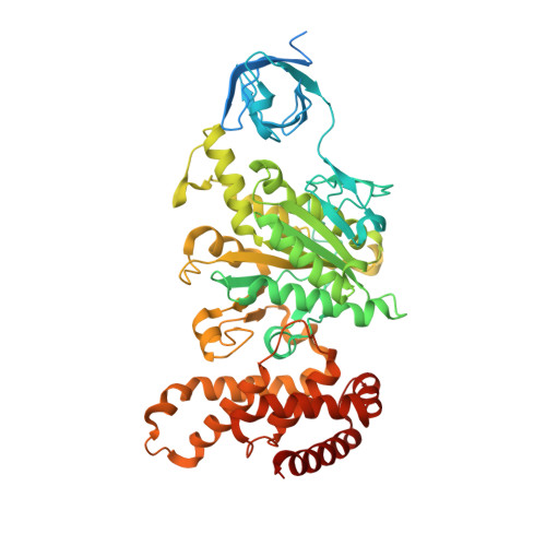
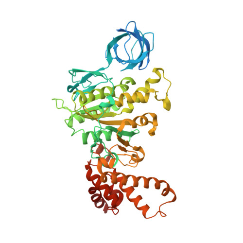
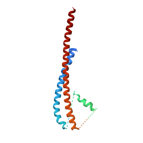
2W6E

Low resolution structures of bovine mitochondrial F1-ATPase during controlled dehydration:hydration state 1.


Experimental Data Snapshot

  • Method: X-RAY DIFFRACTION
  • Resolution: 6.50 Å
  • R-Value Free: 0.285 
  • R-Value Work: 0.290 
  • R-Value Observed: 0.289 

Starting Model: experimental
View more details

wwPDB Validation   3D Report Full Report


Ligand Structure Quality Assessment 


This is version 1.3 of the entry. See complete history


Literature

Improving Diffraction by Humidity Control: A Novel Device Compatible with X-Ray Beamlines.

Sanchez-Weatherby, J.Felisaz, F.Gobbo, A.Huet, J.Ravelli, R.B.G.Bowler, M.W.Cipriani, F.

(2009) Acta Crystallogr D Biol Crystallogr 65: 1237

  • DOI: https://doi.org/10.1107/S0907444909037822
  • Primary Citation of Related Structures:  
    2W6E, 2W6F, 2W6G, 2W6H, 2W6I, 2W6J

  • PubMed Abstract: 

    Dehydration of protein crystals is rarely used, despite being a post-crystallization method that is useful for the improvement of crystal diffraction properties, as it is difficult to reproduce and monitor. A novel device for hydration control of macromolecular crystals in a standard data-collection environment has been developed. The device delivers an air stream of precise relative humidity that can be used to alter the amount of water in macromolecular crystals. The device can be rapidly installed and is fully compatible with most standard synchrotron X-ray beamlines. Samples are mounted in cryoloops and the progress of dehydration can be monitored both optically and by the acquisition of diffraction images. Once the optimal hydration level has been obtained, cryocooling is easy to achieve by hand or by using a sample changer. The device has been thoroughly tested on several ESRF beamlines and is available to users.


  • Organizational Affiliation

    Diamond Light Source Ltd, Diamond House DR1.40, Harwell Science and Innovation Campus, RAL, Chilton, Didcot, Oxfordshire OX11 0DE, England. juan.sanchez-weatherby@diamond.ac.uk


Macromolecules
Find similar proteins by:  (by identity cutoff)  |  3D Structure
Entity ID: 1
MoleculeChains Sequence LengthOrganismDetailsImage
ATP SYNTHASE SUBUNIT ALPHA HEART ISOFORM, MITOCHONDRIAL
A, B, C
553Bos taurusMutation(s): 0 
EC: 3.6.3.14
UniProt
Find proteins for P19483 (Bos taurus)
Explore P19483 
Go to UniProtKB:  P19483
Entity Groups  
Sequence Clusters30% Identity50% Identity70% Identity90% Identity95% Identity100% Identity
UniProt GroupP19483
Sequence Annotations
Expand
  • Reference Sequence
Find similar proteins by:  (by identity cutoff)  |  3D Structure
Entity ID: 2
MoleculeChains Sequence LengthOrganismDetailsImage
ATP SYNTHASE SUBUNIT BETA, MITOCHONDRIAL
D, E, F
528Bos taurusMutation(s): 0 
EC: 3.6.3.14 (PDB Primary Data), 7.1.2.2 (UniProt)
UniProt
Find proteins for P00829 (Bos taurus)
Explore P00829 
Go to UniProtKB:  P00829
Entity Groups  
Sequence Clusters30% Identity50% Identity70% Identity90% Identity95% Identity100% Identity
UniProt GroupP00829
Sequence Annotations
Expand
  • Reference Sequence
Find similar proteins by:  (by identity cutoff)  |  3D Structure
Entity ID: 3
MoleculeChains Sequence LengthOrganismDetailsImage
ATP SYNTHASE SUBUNIT GAMMA, MITOCHONDRIAL298Bos taurusMutation(s): 0 
EC: 3.6.3.14
UniProt
Find proteins for P05631 (Bos taurus)
Explore P05631 
Go to UniProtKB:  P05631
Entity Groups  
Sequence Clusters30% Identity50% Identity70% Identity90% Identity95% Identity100% Identity
UniProt GroupP05631
Sequence Annotations
Expand
  • Reference Sequence
Small Molecules
Ligands 1 Unique
IDChains Name / Formula / InChI Key2D Diagram3D Interactions
ADP
Query on ADP

Download Ideal Coordinates CCD File 
H [auth D]ADENOSINE-5'-DIPHOSPHATE
C10 H15 N5 O10 P2
XTWYTFMLZFPYCI-KQYNXXCUSA-N
Experimental Data & Validation

Experimental Data

  • Method: X-RAY DIFFRACTION
  • Resolution: 6.50 Å
  • R-Value Free: 0.285 
  • R-Value Work: 0.290 
  • R-Value Observed: 0.289 
  • Space Group: P 21 21 21
Unit Cell:
Length ( Å )Angle ( ˚ )
a = 108.49α = 90
b = 141.15β = 90
c = 285.28γ = 90
Software Package:
Software NamePurpose
REFMACrefinement
MOSFLMdata reduction
SCALAdata scaling
MOLREPphasing

Structure Validation

View Full Validation Report


Ligand Structure Quality Assessment 


Entry History 

Deposition Data

Revision History  (Full details and data files)

  • Version 1.0: 2009-10-27
    Type: Initial release
  • Version 1.1: 2011-05-08
    Changes: Version format compliance
  • Version 1.2: 2011-07-13
    Changes: Version format compliance
  • Version 1.3: 2023-12-13
    Changes: Data collection, Database references, Derived calculations, Other, Refinement description