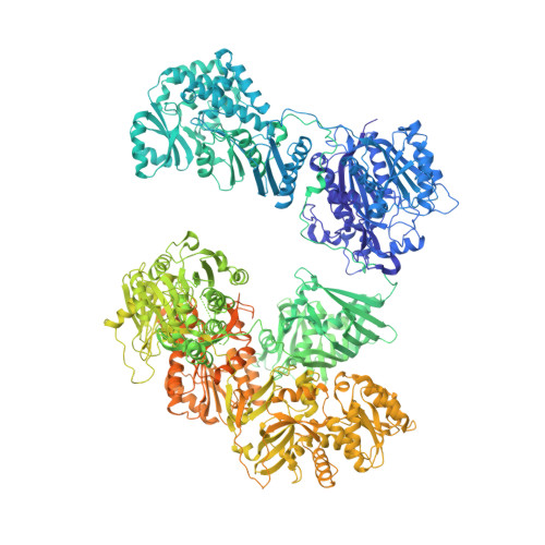
2VZ9

Crystal Structure of Mammalian Fatty Acid Synthase in complex with NADP


Experimental Data Snapshot

  • Method: X-RAY DIFFRACTION
  • Resolution: 3.30 Å
  • R-Value Free: 0.244 
  • R-Value Work: 0.193 

Starting Model: experimental
View more details

wwPDB Validation   3D Report Full Report


Ligand Structure Quality Assessment 


This is version 1.3 of the entry. See complete history


Literature

The Crystal Structure of a Mammalian Fatty Acid Synthase.

Maier, T.Leibundgut, M.Ban, N.

(2008) Science 321: 1315

  • DOI: https://doi.org/10.1126/science.1161269
  • Primary Citation of Related Structures:  
    2VZ8, 2VZ9

  • PubMed Abstract: 

    Mammalian fatty acid synthase is a large multienzyme that catalyzes all steps of fatty acid synthesis. We have determined its crystal structure at 3.2 angstrom resolution covering five catalytic domains, whereas the flexibly tethered terminal acyl carrier protein and thioesterase domains remain unresolved. The structure reveals a complex architecture of alternating linkers and enzymatic domains. Substrate shuttling is facilitated by flexible tethering of the acyl carrier protein domain and by the limited contact between the condensing and modifying portions of the multienzyme, which are mainly connected by linkers rather than direct interaction. The structure identifies two additional nonenzymatic domains: (i) a pseudo-ketoreductase and (ii) a peripheral pseudo-methyltransferase that is probably a remnant of an ancestral methyltransferase domain maintained in some related polyketide synthases. The structural comparison of mammalian fatty acid synthase with modular polyketide synthases shows how their segmental construction allows the variation of domain composition to achieve diverse product synthesis.


  • Organizational Affiliation

    Institute of Molecular Biology and Biophysics, ETH Zurich, 8092 Zurich, Switzerland.


Macromolecules
Find similar proteins by:  (by identity cutoff)  |  3D Structure
Entity ID: 1
MoleculeChains Sequence LengthOrganismDetailsImage
FATTY ACID SYNTHASE
A, B
2,512Sus scrofaMutation(s): 0 
EC: 2.3.1.85 (PDB Primary Data), 2.3.1.41 (UniProt), 3.1.2.14 (UniProt), 1.3.1.39 (UniProt), 2.3.1.38 (UniProt), 4.2.1.59 (UniProt), 1.1.1.100 (UniProt), 2.3.1.39 (UniProt)
UniProt
Find proteins for A5YV76 (Sus scrofa)
Explore A5YV76 
Go to UniProtKB:  A5YV76
Entity Groups  
Sequence Clusters30% Identity50% Identity70% Identity90% Identity95% Identity100% Identity
UniProt GroupA5YV76
Sequence Annotations
Expand
  • Reference Sequence
Experimental Data & Validation

Experimental Data

  • Method: X-RAY DIFFRACTION
  • Resolution: 3.30 Å
  • R-Value Free: 0.244 
  • R-Value Work: 0.193 
  • Space Group: P 1 21 1
Unit Cell:
Length ( Å )Angle ( ˚ )
a = 96.15α = 90
b = 244.89β = 101.84
c = 135.37γ = 90
Software Package:
Software NamePurpose
PHENIXrefinement
XDSdata reduction
SCALAdata scaling

Structure Validation

View Full Validation Report


Ligand Structure Quality Assessment 


Entry History 

Deposition Data

Revision History  (Full details and data files)

  • Version 1.0: 2008-09-09
    Type: Initial release
  • Version 1.1: 2011-05-07
    Changes: Version format compliance
  • Version 1.2: 2011-07-13
    Changes: Version format compliance
  • Version 1.3: 2023-12-13
    Changes: Data collection, Database references, Other, Refinement description