2VVL

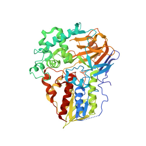
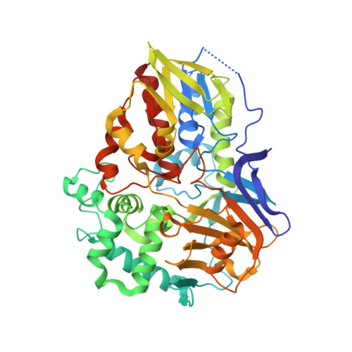
The structure of MAO-N-D3, a variant of monoamine oxidase from Aspergillus niger.


Experimental Data Snapshot

  • Method: X-RAY DIFFRACTION
  • Resolution: 2.45 Å
  • R-Value Free: 0.236 
  • R-Value Work: 0.182 
  • R-Value Observed: 0.185 

Starting Model: experimental
View more details

wwPDB Validation   3D Report Full Report


Ligand Structure Quality Assessment 


This is version 1.3 of the entry. See complete history


Literature

The Structure of Monoamine Oxidase from Aspergillus Niger Provides a Molecular Context for Improvements in Activity Obtained by Directed Evolution.

Atkin, K.E.Reiss, R.Koehler, V.Bailey, K.R.Hart, S.Turkenburg, J.P.Turner, N.J.Brzozowski, A.M.Grogan, G.J.

(2008) J Mol Biol 384: 1218

  • DOI: https://doi.org/10.1016/j.jmb.2008.09.090
  • Primary Citation of Related Structures:  
    2VVL, 2VVM

  • PubMed Abstract: 

    Monoamine oxidase from Aspergillus niger (MAO-N) is a flavoenzyme that catalyses the oxidative deamination of primary amines. MAO-N has been used as the starting model for a series of directed evolution experiments, resulting in mutants of improved activity and broader substrate specificity, suitable for application in the preparative deracemisation of primary, secondary and tertiary amines when used as part of a chemoenzymatic oxidation-reduction cycle. The structures of a three-point mutant (Asn336Ser/Met348Lys/Ile246Met or MAO-N-D3) and a five-point mutant (Asn336Ser/Met348Lys/Ile246Met/Thr384Asn/Asp385Ser or MAO-N-D5) have been obtained using a multiple-wavelength anomalous diffraction experiment on a selenomethionine derivative of the truncated MAO-N-D5 enzyme. MAO-N exists as a homotetramer with a large channel at its centre and shares some structural features with human MAO B (MAO-B). A hydrophobic cavity extends from the protein surface to the active site, where a non-covalently bound flavin adenine dinucleotide (FAD) sits at the base of an 'aromatic cage,' the sides of which are formed by Trp430 and Phe466. A molecule of l-proline was observed near the FAD, and this ligand superimposed well with isatin, a reversible inhibitor of MAO-B, when the structures of MAO-N proline and MAO-B-isatin were overlaid. Of the mutations that confer the ability to catalyse the oxidation of secondary amines in MAO-N-D3, Asn336Ser reduces steric bulk behind Trp430 of the aromatic cage and Ile246Met confers greater flexibility within the substrate binding site. The two additional mutations, Thr384Asn and Asp385Ser, that occur in the MAO-N-D5 variant, which is able to oxidise tertiary amines, appear to influence the active-site environment remotely through changes in tertiary structure that perturb the side chain of Phe382, again altering the steric and electronic character of the active site near FAD. The possible implications of the change in steric and electronic environment caused by relevant mutations are discussed with respect to the improved catalytic efficiency of the MAO-N variants described in the literature.


  • Organizational Affiliation

    York Structural Biology Laboratory, Department of Chemistry, University of York, York YO10 5YW, UK.


Macromolecules
Find similar proteins by:  (by identity cutoff)  |  3D Structure
Entity ID: 1
MoleculeChains Sequence LengthOrganismDetailsImage
MONOAMINE OXIDASE N
A, B, C, D, E
A, B, C, D, E, F, H
495Aspergillus nigerMutation(s): 2 
EC: 1.4.3.4
UniProt
Find proteins for P46882 (Aspergillus niger)
Explore P46882 
Go to UniProtKB:  P46882
Entity Groups  
Sequence Clusters30% Identity50% Identity70% Identity90% Identity95% Identity100% Identity
UniProt GroupP46882
Sequence Annotations
Expand
  • Reference Sequence
Find similar proteins by:  (by identity cutoff)  |  3D Structure
Entity ID: 2
MoleculeChains Sequence LengthOrganismDetailsImage
MONOAMINE OXIDASE N495Aspergillus nigerMutation(s): 2 
EC: 1.4.3.4
UniProt
Find proteins for P46882 (Aspergillus niger)
Explore P46882 
Go to UniProtKB:  P46882
Entity Groups  
Sequence Clusters30% Identity50% Identity70% Identity90% Identity95% Identity100% Identity
UniProt GroupP46882
Sequence Annotations
Expand
  • Reference Sequence
Small Molecules
Ligands 2 Unique
IDChains Name / Formula / InChI Key2D Diagram3D Interactions
FAD
Query on FAD

Download Ideal Coordinates CCD File 
I [auth A]
K [auth B]
L [auth C]
O [auth D]
Q [auth E]
I [auth A],
K [auth B],
L [auth C],
O [auth D],
Q [auth E],
R [auth F],
T [auth G],
W [auth H]
FLAVIN-ADENINE DINUCLEOTIDE
C27 H33 N9 O15 P2
VWWQXMAJTJZDQX-UYBVJOGSSA-N
EDO
Query on EDO

Download Ideal Coordinates CCD File 
J [auth A]
M [auth C]
N [auth C]
P [auth D]
S [auth F]
J [auth A],
M [auth C],
N [auth C],
P [auth D],
S [auth F],
U [auth G],
V [auth G]
1,2-ETHANEDIOL
C2 H6 O2
LYCAIKOWRPUZTN-UHFFFAOYSA-N
Experimental Data & Validation

Experimental Data

  • Method: X-RAY DIFFRACTION
  • Resolution: 2.45 Å
  • R-Value Free: 0.236 
  • R-Value Work: 0.182 
  • R-Value Observed: 0.185 
  • Space Group: P 1 21 1
Unit Cell:
Length ( Å )Angle ( ˚ )
a = 103.287α = 90
b = 187.248β = 90.1
c = 132.432γ = 90
Software Package:
Software NamePurpose
REFMACrefinement
DENZOdata reduction
SCALEPACKdata scaling
MOLREPphasing

Structure Validation

View Full Validation Report


Ligand Structure Quality Assessment 


Entry History 

Deposition Data

Revision History  (Full details and data files)

  • Version 1.0: 2008-11-04
    Type: Initial release
  • Version 1.1: 2011-05-08
    Changes: Version format compliance
  • Version 1.2: 2011-07-13
    Changes: Version format compliance
  • Version 1.3: 2024-05-01
    Changes: Data collection, Database references, Derived calculations, Other, Refinement description