2VUR | pdb_00002vur

Chemical dissection of the link between Streptozotocin, O-GlcNAc and pancreatic cell death


Experimental Data Snapshot

  • Method: X-RAY DIFFRACTION
  • Resolution: 2.20 Å
  • R-Value Free: 
    0.241 (Depositor), 0.197 (DCC) 
  • R-Value Work: 
    0.195 (Depositor), 0.191 (DCC) 
  • R-Value Observed: 
    0.196 (Depositor) 

wwPDB Validation 3D Report Full Report

Validation slider image for 2VUR

Ligand Structure Quality Assessment 


This is version 1.3 of the entry. See complete history

Literature

Chemical Dissection of the Link between Streptozotocin, O-Glcnac, and Pancreatic Cell Death.

Pathak, S.Dorfmueller, H.C.Borodkin, V.S.Van Aalten, D.M.F.

(2008) Chem Biol 15: 799

  • DOI: https://doi.org/10.1016/j.chembiol.2008.06.010
  • Primary Citation Related Structures: 
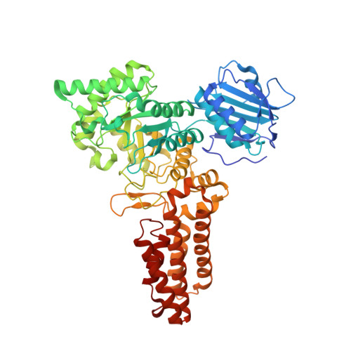
    2VUR

  • PubMed Abstract: 

    Streptozotocin is a natural product that selectively kills insulin-secreting beta cells, and is widely used to generate mouse models of diabetes or treat pancreatic tumors. Several studies suggest that streptozotocin toxicity stems from its N-nitrosourea moiety releasing nitric oxide and possessing DNA alkylating activity. However, it has also been proposed that streptozotocin induces apoptosis by inhibiting O-GlcNAcase, an enzyme that, together with O-GlcNAc transferase, is important for dynamic intracellular protein O-glycosylation. We have used galacto-streptozotocin to chemically dissect the link between O-GlcNAcase inhibition and apoptosis. Using X-ray crystallography, enzymology, and cell biological studies on an insulinoma cell line, we show that, whereas streptozotocin competitively inhibits O-GlcNAcase and induces apoptosis, its galacto-configured derivative no longer inhibits O-GlcNAcase, yet still induces apoptosis. This supports a general chemical poison mode of action for streptozotocin, suggesting the need for using more specific inhibitors to study protein O-GlcNAcylation.


  • Organizational Affiliation
    • Division of Biological Chemistry and Drug Discovery, College of Life Sciences, University of Dundee, Dundee, Scotland.

Macromolecule Content 

  • Total Structure Weight: 134.01 kDa 
  • Atom Count: 9,809 
  • Modeled Residue Count: 1,168 
  • Deposited Residue Count: 1,188 
  • Unique protein chains: 1

Macromolecules

Find similar proteins by:|  3D Structure
Entity ID: 1
MoleculeChains  Sequence LengthOrganismDetailsImage
O-GLCNACASE NAGJ
A, B
594Clostridium perfringensMutation(s): 0 
EC: 3.2.1.35 (PDB Primary Data), 3.2.1.169 (UniProt)
UniProt
Find proteins for Q0TR53 (Clostridium perfringens (strain ATCC 13124 / DSM 756 / JCM 1290 / NCIMB 6125 / NCTC 8237 / Type A))
Explore Q0TR53 
Go to UniProtKB:  Q0TR53
Entity Groups
Sequence Clusters30% Identity50% Identity70% Identity90% Identity95% Identity100% Identity
UniProt GroupQ0TR53
Sequence Annotations
Expand
Reference Sequence

Experimental Data & Validation

Experimental Data

  • Method: X-RAY DIFFRACTION
  • Resolution: 2.20 Å
  • R-Value Free:  0.241 (Depositor), 0.197 (DCC) 
  • R-Value Work:  0.195 (Depositor), 0.191 (DCC) 
  • R-Value Observed: 0.196 (Depositor) 
Space Group: I 21 21 21
Unit Cell:
Length ( Å )Angle ( ˚ )
a = 130.47α = 90
b = 149.731β = 90
c = 152.121γ = 90
Software Package:
Software NamePurpose
REFMACrefinement
DENZOdata reduction
SCALEPACKdata scaling
MOLREPphasing

Structure Validation

View Full Validation Report


Ligand Structure Quality Assessment 


Entry History 

Deposition Data

Revision History  (Full details and data files)

  • Version 1.0: 2009-02-10
    Type: Initial release
  • Version 1.1: 2011-07-13
    Changes: Advisory, Version format compliance
  • Version 1.2: 2020-07-29
    Type: Remediation
    Reason: Carbohydrate remediation
    Changes: Data collection, Derived calculations, Other
  • Version 1.3: 2024-05-08
    Changes: Data collection, Database references, Structure summary