2VTB | pdb_00002vtb

Structure of cryptochrome 3 - DNA complex


Experimental Data Snapshot

  • Method: X-RAY DIFFRACTION
  • Resolution: 2.01 Å
  • R-Value Free: 
    0.222 (Depositor), 0.231 (DCC) 
  • R-Value Work: 
    0.184 (Depositor), 0.195 (DCC) 
  • R-Value Observed: 
    0.185 (Depositor) 

Starting Model: experimental
View more details

wwPDB Validation 3D Report Full Report

Validation slider image for 2VTB

Ligand Structure Quality Assessment 


This is version 1.3 of the entry. See complete history

Literature

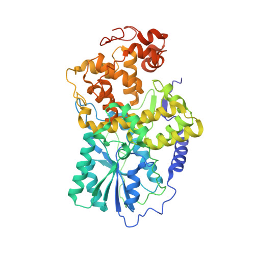
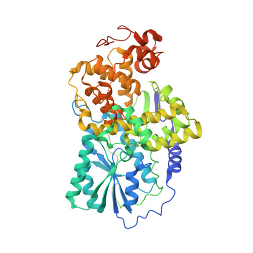
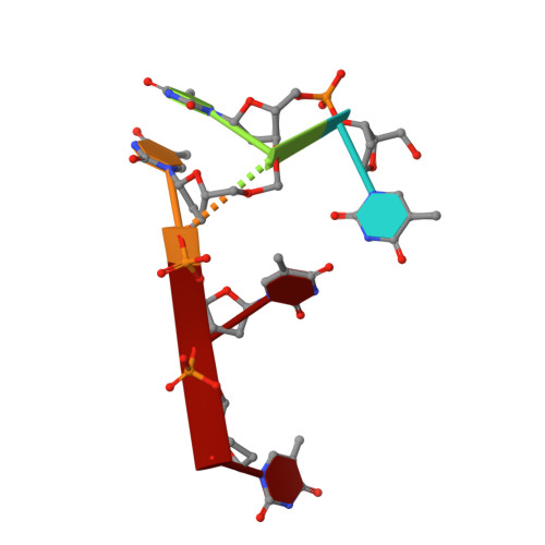
Recognition and Repair of Uv Lesions in Loop Structures of Duplex DNA by Dash-Type Cryptochrome.

Pokorny, R.Klar, T.Hennecke, U.Carell, T.Batschauer, A.Essen, L.-O.

(2008) Proc Natl Acad Sci U S A 105: 21023

  • DOI: https://doi.org/10.1073/pnas.0805830106
  • Primary Citation Related Structures: 
    2VTB

  • PubMed Abstract: 

    DNA photolyases and cryptochromes (cry) form a family of flavoproteins that use light energy in the blue/UV-A region for the repair of UV-induced DNA lesions or for signaling, respectively. Very recently, it was shown that members of the DASH cryptochrome subclade repair specifically cyclobutane pyrimidine dimers (CPDs) in UV-damaged single-stranded DNA. Here, we report the crystal structure of Arabidopsis cryptochrome 3 with an in-situ-repaired CPD substrate in single-stranded DNA. The structure shows a binding mode similar to that of conventional DNA photolyases. Furthermore, CPD lesions in double-stranded DNA are bound and repaired with similar efficiency as in single-stranded DNA if the CPD lesion is present in a loop structure. Together, these data reveal that DASH cryptochromes catalyze light-driven DNA repair like conventional photolyases but lack an efficient flipping mechanism for interaction with CPD lesions within duplex DNA.


  • Organizational Affiliation
    • Department of Biology, Philipps University, Karl-von-Frisch-Strasse 8, D-35032 Marburg, Germany.

Macromolecule Content 

  • Total Structure Weight: 378.97 kDa 
  • Atom Count: 26,329 
  • Modeled Residue Count: 2,974 
  • Deposited Residue Count: 3,185 
  • Unique protein chains: 2
  • Unique nucleic acid chains: 1

Macromolecules


Find similar proteins by:|  3D Structure
Entity ID: 1
MoleculeChains  Sequence LengthOrganismDetailsImage
CRYPTOCHROME DASH
A, C, D, E, F
526Arabidopsis thalianaMutation(s): 0 
EC: 4.1.99.3
UniProt
Find proteins for Q84KJ5 (Arabidopsis thaliana)
Explore Q84KJ5 
Go to UniProtKB:  Q84KJ5
Entity Groups
Sequence Clusters30% Identity50% Identity70% Identity90% Identity95% Identity100% Identity
UniProt GroupQ84KJ5
Sequence Annotations
Expand
Reference Sequence
Find similar proteins by:|  3D Structure
Entity ID: 2
MoleculeChains  Sequence LengthOrganismDetailsImage
CRYPTOCHROME DASH525Arabidopsis thalianaMutation(s): 0 
EC: 4.1.99.3
UniProt
Find proteins for Q84KJ5 (Arabidopsis thaliana)
Explore Q84KJ5 
Go to UniProtKB:  Q84KJ5
Entity Groups
Sequence Clusters30% Identity50% Identity70% Identity90% Identity95% Identity100% Identity
UniProt GroupQ84KJ5
Sequence Annotations
Expand
Reference Sequence
Find similar nucleic acids by:  Sequence
Entity ID: 3
MoleculeChains LengthOrganismImage
5'-D(*DT*DT*DT*DT*DTP)-3'
G, H, I, J, K
G, H, I, J, K, L
5Homo sapiens
Sequence Annotations
Expand
Reference Sequence

Small Molecules

Ligands 4 Unique
IDChains Name / Formula / InChI Key2D Diagram3D Interactions
FAD

Query on FAD


Download:Ideal Coordinates CCD File
BA [auth E]
EA [auth F]
M [auth A]
P [auth B]
U [auth C]
BA [auth E],
EA [auth F],
M [auth A],
P [auth B],
U [auth C],
Y [auth D]
FLAVIN-ADENINE DINUCLEOTIDE
C27 H33 N9 O15 P2
VWWQXMAJTJZDQX-UYBVJOGSSA-N
MHF

Query on MHF


Download:Ideal Coordinates CCD File
CA [auth E]
FA [auth F]
N [auth A]
Q [auth B]
V [auth C]
CA [auth E],
FA [auth F],
N [auth A],
Q [auth B],
V [auth C],
Z [auth D]
5,10-METHENYL-6,7,8-TRIHYDROFOLIC ACID
C20 H23 N7 O6
QYNUQALWYRSVHF-OLZOCXBDSA-N
ACT

Query on ACT


Download:Ideal Coordinates CCD File
T [auth B]ACETATE ION
C2 H3 O2
QTBSBXVTEAMEQO-UHFFFAOYSA-M
CL

Query on CL


Download:Ideal Coordinates CCD File
AA [auth D]
DA [auth E]
GA [auth F]
O [auth A]
R [auth B]
AA [auth D],
DA [auth E],
GA [auth F],
O [auth A],
R [auth B],
S [auth B],
W [auth C],
X [auth C]
CHLORIDE ION
Cl
VEXZGXHMUGYJMC-UHFFFAOYSA-M

Experimental Data & Validation

Experimental Data

  • Method: X-RAY DIFFRACTION
  • Resolution: 2.01 Å
  • R-Value Free:  0.222 (Depositor), 0.231 (DCC) 
  • R-Value Work:  0.184 (Depositor), 0.195 (DCC) 
  • R-Value Observed: 0.185 (Depositor) 
Space Group: P 21 21 21
Unit Cell:
Length ( Å )Angle ( ˚ )
a = 120.734α = 90
b = 136.082β = 90
c = 211.484γ = 90
Software Package:
Software NamePurpose
REFMACrefinement
MOSFLMdata reduction
SCALAdata scaling
PHASERphasing

Structure Validation

View Full Validation Report


Ligand Structure Quality Assessment 


Entry History 

Deposition Data

Revision History  (Full details and data files)

  • Version 1.0: 2009-06-02
    Type: Initial release
  • Version 1.1: 2011-07-13
    Changes: Advisory, Version format compliance
  • Version 1.2: 2019-07-24
    Changes: Advisory, Data collection, Derived calculations
  • Version 1.3: 2023-12-13
    Changes: Advisory, Data collection, Database references, Derived calculations, Other, Refinement description