2VDB | pdb_00002vdb

Structure of human serum albumin with S-naproxen and the GA module


Experimental Data Snapshot

  • Method: X-RAY DIFFRACTION
  • Resolution: 2.52 Å
  • R-Value Free: 
    0.280 (Depositor), 0.263 (DCC) 
  • R-Value Work: 
    0.234 (Depositor), 0.224 (DCC) 
  • R-Value Observed: 
    0.236 (Depositor) 

Starting Models: experimental
View more details

wwPDB Validation 3D Report Full Report

Validation slider image for 2VDB

Ligand Structure Quality Assessment 


This is version 1.3 of the entry. See complete history

Literature

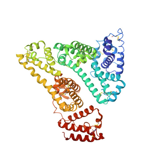
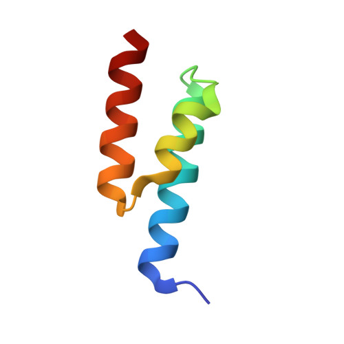
Structural Basis for the Binding of Naproxen to Human Serum Albumin in the Presence of Fatty Acids and the Ga Module.

Lejon, S.Cramer, J.F.Nordberg, P.A.

(2008) Acta Crystallogr Sect F Struct Biol Cryst Commun 64: 64

  • DOI: https://doi.org/10.1107/S174430910706770X
  • Primary Citation Related Structures: 
    2VDB

  • PubMed Abstract: 

    The previously determined crystal structure of the bacterial albumin-binding GA module in complex with human serum albumin (HSA) suggested the possibility of utilizing the complex in the study of ligand binding to HSA. As a continuation of these studies, the crystal structure of the HSA-GA complex with the drug molecule naproxen and the fatty acid decanoate bound to HSA has been determined to a resolution of 2.5 A. In terms of drug binding, the structure suggests that the binding of decanoate to the albumin molecule may play a role in making the haemin site in subdomain IB of the albumin molecule available for the binding of naproxen. In addition, structure comparisons with solved structures of HSA and of the HSA-GA complex show that the GA module is capable of binding to different conformations of HSA. The HSA-GA complex therefore emerges as a possible platform for the crystallographic study of specific HSA-drug interactions and of the influence exerted by the presence of fatty acids.


  • Organizational Affiliation
    • Department of Cell and Molecular Biology, Uppsala University, Biomedical Centre, Box 596, S-751 24 Uppsala, Sweden. sara@xray.bmc.uu.se

Macromolecule Content 

  • Total Structure Weight: 73.39 kDa 
  • Atom Count: 4,872 
  • Modeled Residue Count: 629 
  • Deposited Residue Count: 634 
  • Unique protein chains: 2

Macromolecules

Find similar proteins by:|  3D Structure
Entity ID: 1
MoleculeChains  Sequence LengthOrganismDetailsImage
SERUM ALBUMIN579Homo sapiensMutation(s): 0 
UniProt & NIH Common Fund Data Resources
Find proteins for P02768 (Homo sapiens)
Explore P02768 
Go to UniProtKB:  P02768
PHAROS:  P02768
GTEx:  ENSG00000163631 
Entity Groups
Sequence Clusters30% Identity50% Identity70% Identity90% Identity95% Identity100% Identity
UniProt GroupP02768
Sequence Annotations
Expand
Reference Sequence
Find similar proteins by:|  3D Structure
Entity ID: 2
MoleculeChains  Sequence LengthOrganismDetailsImage
PEPTOSTREPTOCOCCAL ALBUMIN-BINDING PROTEIN55Finegoldia magnaMutation(s): 0 
UniProt
Find proteins for Q51911 (Finegoldia magna)
Explore Q51911 
Go to UniProtKB:  Q51911
Entity Groups
Sequence Clusters30% Identity50% Identity70% Identity90% Identity95% Identity100% Identity
UniProt GroupQ51911
Sequence Annotations
Expand
Reference Sequence

Small Molecules

Experimental Data & Validation

Experimental Data

  • Method: X-RAY DIFFRACTION
  • Resolution: 2.52 Å
  • R-Value Free:  0.280 (Depositor), 0.263 (DCC) 
  • R-Value Work:  0.234 (Depositor), 0.224 (DCC) 
  • R-Value Observed: 0.236 (Depositor) 
Space Group: C 1 2 1
Unit Cell:
Length ( Å )Angle ( ˚ )
a = 190.507α = 90
b = 49.462β = 93
c = 79.933γ = 90
Software Package:
Software NamePurpose
REFMACrefinement
MOSFLMdata reduction
SCALAdata scaling
TRUNCATEdata scaling
MOLREPphasing

Structure Validation

View Full Validation Report


Ligand Structure Quality Assessment 


Entry History 

Deposition Data

Revision History  (Full details and data files)

  • Version 1.0: 2008-02-26
    Type: Initial release
  • Version 1.1: 2011-07-13
    Changes: Advisory, Version format compliance
  • Version 1.2: 2023-12-13
    Changes: Data collection, Database references, Other, Refinement description
  • Version 1.3: 2024-10-23
    Changes: Structure summary