2VBQ | pdb_00002vbq

Structure of AAC(6')-Iy in complex with bisubstrate analog CoA-S- monomethyl-acetylneamine.


Experimental Data Snapshot

  • Method: X-RAY DIFFRACTION
  • Resolution: 2.00 Å
  • R-Value Free: 
    0.230 (Depositor), 0.228 (DCC) 
  • R-Value Work: 
    0.181 (Depositor), 0.180 (DCC) 
  • R-Value Observed: 
    0.183 (Depositor) 

wwPDB Validation 3D Report Full Report

Validation slider image for 2VBQ

Ligand Structure Quality Assessment 


This is version 1.6 of the entry. See complete history

Literature

Kinetic and Structural Analysis of Bisubstrate Inhibition of the Salmonella Enterica Aminoglycoside 6'-N-Acetyltransferase.

Magalhaes, M.L.Vetting, M.W.Gao, F.Freiburger, L.Auclair, K.Blanchard, J.S.

(2008) Biochemistry 47: 579

  • DOI: https://doi.org/10.1021/bi701957c
  • Primary Citation Related Structures: 
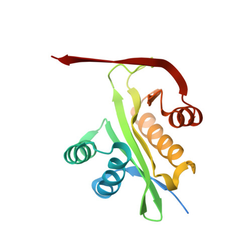
    2VBQ

  • PubMed Abstract: 

    Aminoglycosides are antibacterial compounds that act by binding to the A site of the small 30S bacterial ribosomal subunit and inhibiting protein translation. Clinical resistance to aminoglycosides is generally the result of the expression of enzymes that covalently modify the antibiotic, including phosphorylation, adenylylation, and acetylation. Bisubstrate analogs for the aminoglycoside N-acetyltransferases are nanomolar inhibitors of Enterococcus faecium AAC(6')-Ii. However, in the case of the Salmonella enterica aac(6')-Iy-encoded aminoglycoside N-acetyltransferase, we demonstrate that a series of bisubstrate analogs are only micromolar inhibitors. In contrast to studies with AAC(6')-Ii, the inhibition constants toward AAC(6')-Iy are essentially independent of both the identity of the aminoglycoside component of the bisubstrate and the number of carbon atoms that are used to link the CoA and aminoglycoside components. The patterns of inhibition suggest that the CoA portion of the bisubstrate analog can bind to the enzyme-aminoglycoside substrate complex and that the aminoglycoside portion can bind to the enzyme-CoA product complex. However, at the high concentrations of bisubstrate analog used in crystallization experiments, we could crystallize and solve the three-dimensional structure of the enzyme-bisubstrate complex. The structure reveals that both the CoA and aminoglycoside portions bind in essentially the same positions as those previously observed for the enzyme-CoA-ribostamycin complex, with only a modest adjustment to accommodate the "linker". These results are compared to previous studies of the interaction of similar bisubstrate analogs with other aminoglycoside N-acetyltransferases.


  • Organizational Affiliation
    • Department of Biochemistry, Albert Einstein College of Medicine, 1300 Morris Park Avenue, Bronx, New York 10461, USA.

Macromolecule Content 

  • Total Structure Weight: 39.55 kDa 
  • Atom Count: 2,684 
  • Modeled Residue Count: 291 
  • Deposited Residue Count: 330 
  • Unique protein chains: 1

Macromolecules

Find similar proteins by:|  3D Structure
Entity ID: 1
MoleculeChains  Sequence LengthOrganismDetailsImage
AMINOGLYCOSIDE 6'-N-ACETYLTRANSFERASE
A, B
165Salmonella entericaMutation(s): 0 
EC: 2.3.1.82
UniProt
Find proteins for Q9R381 (Salmonella enteritidis)
Explore Q9R381 
Go to UniProtKB:  Q9R381
Entity Groups
Sequence Clusters30% Identity50% Identity70% Identity90% Identity95% Identity100% Identity
UniProt GroupQ9R381
Sequence Annotations
Expand
Reference Sequence

Small Molecules

Ligands 3 Unique
IDChains Name / Formula / InChI Key2D Diagram3D Interactions
BSJ

Query on BSJ


Download:Ideal Coordinates CCD File
C [auth A],
F [auth B]
(3R,9Z)-17-[(2R,3S,4R,5R,6R)-5-amino-6-{[(1R,2R,3S,4R,6S)-4,6-diamino-2,3-dihydroxycyclohexyl]oxy}-3,4-dihydroxytetrahydro-2H-pyran-2-yl]-3-hydroxy-2,2-dimethyl-4,8,15-trioxo-12-thia-5,9,16-triazaheptadec-9-en-1-yl [(2R,3S,4R,5R)-5-(6-amino-9H-purin-9-yl)-4-hydroxy-3-(phosphonooxy)tetrahydrofuran-2-yl]methyl dihydrogen diphosphate
C36 H62 N11 O23 P3 S
HXXBCMJRHXNFMC-BOUWNZBBSA-N
GOL

Query on GOL


Download:Ideal Coordinates CCD File
E [auth A]GLYCEROL
C3 H8 O3
PEDCQBHIVMGVHV-UHFFFAOYSA-N
NI

Query on NI


Download:Ideal Coordinates CCD File
D [auth A]NICKEL (II) ION
Ni
VEQPNABPJHWNSG-UHFFFAOYSA-N

Experimental Data & Validation

Experimental Data

  • Method: X-RAY DIFFRACTION
  • Resolution: 2.00 Å
  • R-Value Free:  0.230 (Depositor), 0.228 (DCC) 
  • R-Value Work:  0.181 (Depositor), 0.180 (DCC) 
  • R-Value Observed: 0.183 (Depositor) 
Space Group: C 1 2 1
Unit Cell:
Length ( Å )Angle ( ˚ )
a = 84.964α = 90
b = 44.599β = 93.92
c = 88.387γ = 90
Software Package:
Software NamePurpose
REFMACrefinement
MOSFLMdata reduction
SCALAdata scaling
REFMACphasing

Structure Validation

View Full Validation Report


Ligand Structure Quality Assessment 


Entry History 

Deposition Data

Revision History  (Full details and data files)

  • Version 1.0: 2008-01-08
    Type: Initial release
  • Version 1.1: 2011-05-08
    Changes: Version format compliance
  • Version 1.2: 2011-07-13
    Changes: Version format compliance
  • Version 1.3: 2017-07-05
    Changes: Data collection
  • Version 1.4: 2019-03-06
    Changes: Data collection, Experimental preparation
  • Version 1.5: 2020-03-11
    Changes: Derived calculations, Other
  • Version 1.6: 2024-05-08
    Changes: Data collection, Database references, Derived calculations