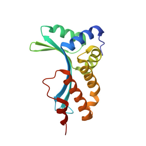
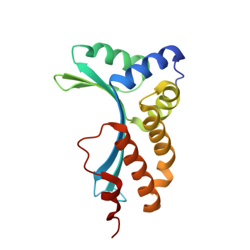
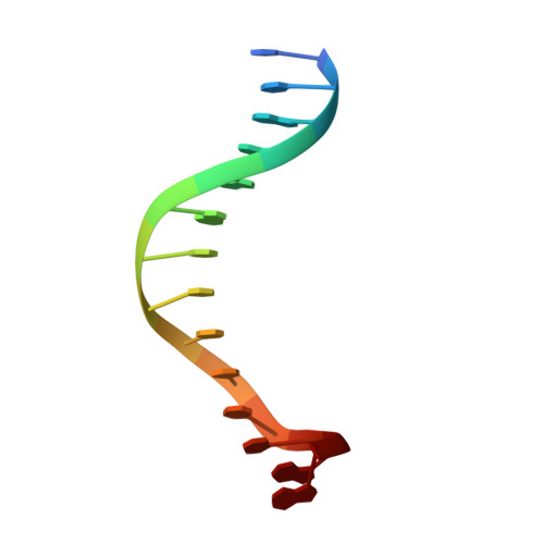
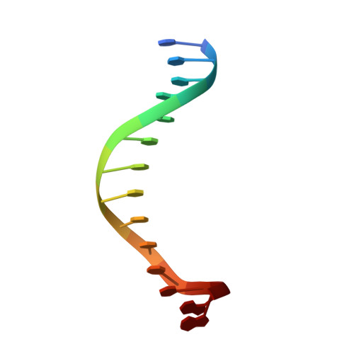
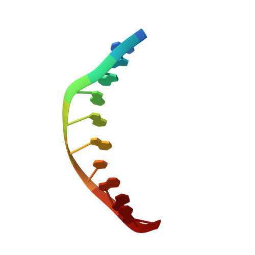
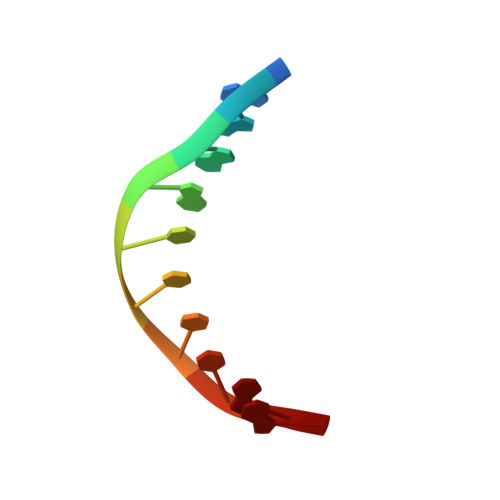
2VBN

Molecular basis of human XPC gene recognition and cleavage by engineered homing endonuclease heterodimers


Experimental Data Snapshot

  • Method: X-RAY DIFFRACTION
  • Resolution: 1.90 Å
  • R-Value Free: 0.223 
  • R-Value Work: 0.141 
  • R-Value Observed: 0.145 

Starting Model: experimental
View more details

wwPDB Validation   3D Report Full Report


This is version 1.2 of the entry. See complete history


Literature

Molecular Basis of Xeroderma Pigmentosum Group C DNA Recognition by Engineered Meganucleases

Redondo, P.Prieto, J.Munoz, I.G.Alibes, A.Stricher, F.Serrano, L.Cabaniols, J.P.Daboussi, F.Arnould, S.Perez, C.Duchateau, P.Paques, F.Blanco, F.J.Montoya, G.

(2008) Nature 456: 107

  • DOI: https://doi.org/10.1038/nature07343
  • Primary Citation of Related Structures:  
    2VBJ, 2VBL, 2VBN, 2VBO

  • PubMed Abstract: 

    Xeroderma pigmentosum is a monogenic disease characterized by hypersensitivity to ultraviolet light. The cells of xeroderma pigmentosum patients are defective in nucleotide excision repair, limiting their capacity to eliminate ultraviolet-induced DNA damage, and resulting in a strong predisposition to develop skin cancers. The use of rare cutting DNA endonucleases-such as homing endonucleases, also known as meganucleases-constitutes one possible strategy for repairing DNA lesions. Homing endonucleases have emerged as highly specific molecular scalpels that recognize and cleave DNA sites, promoting efficient homologous gene targeting through double-strand-break-induced homologous recombination. Here we describe two engineered heterodimeric derivatives of the homing endonuclease I-CreI, produced by a semi-rational approach. These two molecules-Amel3-Amel4 and Ini3-Ini4-cleave DNA from the human XPC gene (xeroderma pigmentosum group C), in vitro and in vivo. Crystal structures of the I-CreI variants complexed with intact and cleaved XPC target DNA suggest that the mechanism of DNA recognition and cleavage by the engineered homing endonucleases is similar to that of the wild-type I-CreI. Furthermore, these derivatives induced high levels of specific gene targeting in mammalian cells while displaying no obvious genotoxicity. Thus, homing endonucleases can be designed to recognize and cleave the DNA sequences of specific genes, opening up new possibilities for genome engineering and gene therapy in xeroderma pigmentosum patients whose illness can be treated ex vivo.


  • Organizational Affiliation

    Macromolecular Crystallography Group, Spanish National Cancer Research Centre (CNIO), c/Melchor Fdez. Almagro 3, 28029 Madrid, Spain.


Macromolecules

Find similar proteins by:  (by identity cutoff)  |  3D Structure
Entity ID: 1
MoleculeChains Sequence LengthOrganismDetailsImage
DNA ENDONUCLEASE I-CREI153Chlamydomonas reinhardtiiMutation(s): 0 
EC: 3.1
UniProt
Find proteins for P05725 (Chlamydomonas reinhardtii)
Explore P05725 
Go to UniProtKB:  P05725
Entity Groups  
Sequence Clusters30% Identity50% Identity70% Identity90% Identity95% Identity100% Identity
UniProt GroupP05725
Sequence Annotations
Expand
  • Reference Sequence
Find similar proteins by:  (by identity cutoff)  |  3D Structure
Entity ID: 2
MoleculeChains Sequence LengthOrganismDetailsImage
DNA ENDONUCLEASE I-CREI153Chlamydomonas reinhardtiiMutation(s): 0 
EC: 3.1
UniProt
Find proteins for P05725 (Chlamydomonas reinhardtii)
Explore P05725 
Go to UniProtKB:  P05725
Entity Groups  
Sequence Clusters30% Identity50% Identity70% Identity90% Identity95% Identity100% Identity
UniProt GroupP05725
Sequence Annotations
Expand
  • Reference Sequence

Find similar nucleic acids by:  Sequence   |   3D Structure  

Entity ID: 3
MoleculeChains LengthOrganismImage
5'-D(*TP*TP*AP*GP*GP*AP*TP*CP*CP*TP *TP*CP*AP*AP)-3'14N/A
Sequence Annotations
Expand
  • Reference Sequence

Find similar nucleic acids by:  Sequence   |   3D Structure  

Entity ID: 4
MoleculeChains LengthOrganismImage
5'-D(*TP*CP*TP*GP*CP*CP*TP*TP*TP*TP *TP*TP*GP*AP)-3'D [auth E]14N/A
Sequence Annotations
Expand
  • Reference Sequence

Find similar nucleic acids by:  Sequence   |   3D Structure  

Entity ID: 5
MoleculeChains LengthOrganismImage
5'-D(*AP*AP*AP*AP*GP*GP*CP*AP*GP*AP)-3'E [auth S]10N/A
Sequence Annotations
Expand
  • Reference Sequence

Find similar nucleic acids by:  Sequence   |   3D Structure  

Entity ID: 6
MoleculeChains LengthOrganismImage
5'-D(*AP*GP*GP*AP*TP*CP*CP*TP*AP*AP)-3'F [auth T]10N/A
Sequence Annotations
Expand
  • Reference Sequence
Experimental Data & Validation

Experimental Data

  • Method: X-RAY DIFFRACTION
  • Resolution: 1.90 Å
  • R-Value Free: 0.223 
  • R-Value Work: 0.141 
  • R-Value Observed: 0.145 
  • Space Group: P 1 21 1
Unit Cell:
Length ( Å )Angle ( ˚ )
a = 46.115α = 90
b = 67.992β = 90.11
c = 77.186γ = 90
Software Package:
Software NamePurpose
REFMACrefinement
HKL-2000data reduction
SCALEPACKdata scaling
PHASERphasing

Structure Validation

View Full Validation Report


Entry History 

Revision History  (Full details and data files)

  • Version 1.0: 2008-10-28
    Type: Initial release
  • Version 1.1: 2012-06-20
    Changes: Database references, Derived calculations, Non-polymer description, Other, Version format compliance
  • Version 1.2: 2023-12-13
    Changes: Data collection, Database references, Derived calculations, Other, Refinement description