2VBF | pdb_00002vbf

The holostructure of the branched-chain keto acid decarboxylase (KdcA) from Lactococcus lactis


Experimental Data Snapshot

  • Method: X-RAY DIFFRACTION
  • Resolution: 1.60 Å
  • R-Value Free: 
    0.200 (Depositor), 0.206 (DCC) 
  • R-Value Work: 
    0.163 (Depositor), 0.170 (DCC) 
  • R-Value Observed: 
    0.165 (Depositor) 

Starting Model: experimental
View more details

wwPDB Validation 3D Report Full Report

Validation slider image for 2VBF

Ligand Structure Quality Assessment 


This is version 1.3 of the entry. See complete history

Literature

Crystal Structure of the Branched-Chain Keto Acid Decarboxylase (Kdca) from Lactococcus Lactis Provides Insights Into the Structural Basis for the Chemo- and Enantioselective Carboligation Reaction

Berthold, C.L.Gocke, D.Wood, M.D.Leeper, F.Pohl, M.Schneider, G.

(2007) Acta Crystallogr D Biol Crystallogr 63: 1217

  • DOI: https://doi.org/10.1107/S0907444907050433
  • Primary Citation Related Structures: 
    2VBF, 2VBG

  • PubMed Abstract: 

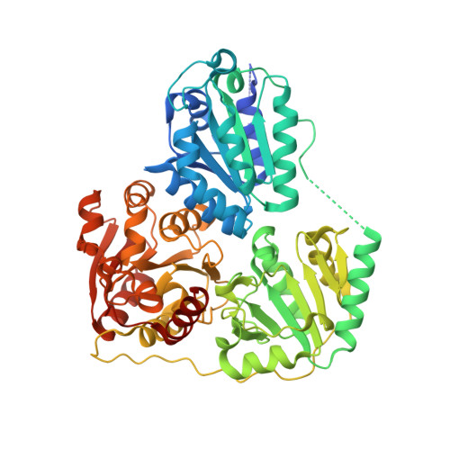
    The thiamin diphosphate (ThDP) dependent branched-chain keto acid decarboxylase (KdcA) from Lactococcus lactis catalyzes the decarboxylation of 3-methyl-2-oxobutanoic acid to 3-methylpropanal (isobutyraldehyde) and CO2. The enzyme is also able to catalyze carboligation reactions with an exceptionally broad substrate range, a feature that makes KdcA a potentially valuable biocatalyst for C-C bond formation, in particular for the enzymatic synthesis of diversely substituted 2-hydroxyketones with high enantioselectivity. The crystal structures of recombinant holo-KdcA and of a complex with an inhibitory ThDP analogue mimicking a reaction intermediate have been determined to resolutions of 1.6 and 1.8 A, respectively. KdcA shows the fold and cofactor-protein interactions typical of thiamin-dependent enzymes. In contrast to the tetrameric assembly displayed by most other ThDP-dependent decarboxylases of known structure, KdcA is a homodimer. The crystal structures provide insights into the structural basis of substrate selectivity and stereoselectivity of the enzyme and thus are suitable as a framework for the redesign of the substrate profile in carboligation reactions.


  • Organizational Affiliation
    • Department of Medical Biochemistry and Biophysics, Karolinska Institutet, S-17177 Stockholm, Sweden.

Macromolecule Content 

  • Total Structure Weight: 127.71 kDa 
  • Atom Count: 9,807 
  • Modeled Residue Count: 1,092 
  • Deposited Residue Count: 1,140 
  • Unique protein chains: 1

Macromolecules

Find similar proteins by:|  3D Structure
Entity ID: 1
MoleculeChains  Sequence LengthOrganismDetailsImage
BRANCHED-CHAIN ALPHA-KETOACID DECARBOXYLASE
A, B
570Lactococcus lactisMutation(s): 0 
UniProt
Find proteins for Q6QBS4 (Lactococcus lactis)
Explore Q6QBS4 
Go to UniProtKB:  Q6QBS4
Entity Groups
Sequence Clusters30% Identity50% Identity70% Identity90% Identity95% Identity100% Identity
UniProt GroupQ6QBS4
Sequence Annotations
Expand
Reference Sequence

Experimental Data & Validation

Experimental Data

  • Method: X-RAY DIFFRACTION
  • Resolution: 1.60 Å
  • R-Value Free:  0.200 (Depositor), 0.206 (DCC) 
  • R-Value Work:  0.163 (Depositor), 0.170 (DCC) 
  • R-Value Observed: 0.165 (Depositor) 
Space Group: P 21 21 21
Unit Cell:
Length ( Å )Angle ( ˚ )
a = 65.603α = 90
b = 108.421β = 90
c = 146.593γ = 90
Software Package:
Software NamePurpose
REFMACrefinement
MOSFLMdata reduction
SCALAdata scaling
MOLREPphasing

Structure Validation

View Full Validation Report


Ligand Structure Quality Assessment 


Entry History 

Deposition Data

Revision History  (Full details and data files)

  • Version 1.0: 2007-12-18
    Type: Initial release
  • Version 1.1: 2011-05-08
    Changes: Version format compliance
  • Version 1.2: 2011-07-13
    Changes: Version format compliance
  • Version 1.3: 2023-12-13
    Changes: Data collection, Database references, Derived calculations, Other, Refinement description