2VA2

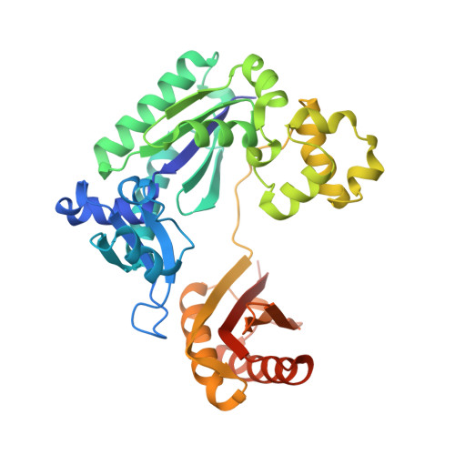
Complex structure of Sulfolobus solfataricus DPO4 and DNA duplex containing a hydrophobic thymine isostere 2,4-difluorotoluene nucleotide in the template strand


Experimental Data Snapshot

  • Method: X-RAY DIFFRACTION
  • Resolution: 2.80 Å
  • R-Value Free: 0.250 
  • R-Value Work: 0.211 
  • R-Value Observed: 0.211 

Starting Model: experimental
View more details

wwPDB Validation   3D Report Full Report


Ligand Structure Quality Assessment 


This is version 1.2 of the entry. See complete history


Literature

Structure and Activity of Y-Class DNA Polymerase Dpo4 from Sulfolobus Solfataricus with Templates Containing the Hydrophobic Thymine Analog 2,4- Difluorotoluene.

Irimia, A.Eoff, R.L.Pallan, P.S.Guengerich, F.P.Egli, M.

(2007) J Biol Chem 282: 36421

  • DOI: https://doi.org/10.1074/jbc.M707267200
  • Primary Citation of Related Structures:  
    2V9W, 2VA2, 2VA3

  • PubMed Abstract: 

    The 2,4-difluorotoluene (DFT) analog of thymine has been used extensively to probe the relative importance of shape and hydrogen bonding for correct nucleotide insertion by DNA polymerases. As far as high fidelity (A-class) polymerases are concerned, shape is considered by some as key to incorporation of A(T) opposite T(A) and G(C) opposite C(G). We have carried out a detailed kinetic analysis of in vitro primer extension opposite DFT-containing templates by the trans-lesion (Y-class) DNA polymerase Dpo4 from Sulfolobus solfataricus. Although full-length product formation was observed, steady-state kinetic data show that dATP insertion opposite DFT is greatly inhibited relative to insertion opposite T (approximately 5,000-fold). No products were observed in the pre-steady-state. Furthermore, it is noteworthy that Dpo4 strongly prefers dATP opposite DFT over dGTP (approximately 200-fold) and that the polymerase is able to extend an A:DFT but not a G:DFT pair. We present crystal structures of Dpo4 in complex with DNA duplexes containing the DFT analog, the first for any DNA polymerase. In the structures, template-DFT is either positioned opposite primer-A or -G at the -1 site or is unopposed by a primer base and followed by a dGTP:A mismatch pair at the active site, representative of a -1 frameshift. The three structures provide insight into the discrimination by Dpo4 between dATP and dGTP opposite DFT and its inability to extend beyond a G:DFT pair. Although hydrogen bonding is clearly important for error-free replication by this Y-class DNA polymerase, our work demonstrates that Dpo4 also relies on shape and electrostatics to distinguish between correct and incorrect incoming nucleotide.


  • Organizational Affiliation

    Department of Biochemistry and Center in Molecular Toxicology, Vanderbilt University School of Medicine, Nashville, Tennessee 37232-0146, USA.


Macromolecules

Find similar proteins by:  (by identity cutoff)  |  3D Structure
Entity ID: 1
MoleculeChains Sequence LengthOrganismDetailsImage
DNA POLYMERASE IV
A, B
358Saccharolobus solfataricus P2Mutation(s): 0 
EC: 2.7.7.7
UniProt
Find proteins for Q97W02 (Saccharolobus solfataricus (strain ATCC 35092 / DSM 1617 / JCM 11322 / P2))
Explore Q97W02 
Go to UniProtKB:  Q97W02
Entity Groups  
Sequence Clusters30% Identity50% Identity70% Identity90% Identity95% Identity100% Identity
UniProt GroupQ97W02
Sequence Annotations
Expand
  • Reference Sequence

Find similar nucleic acids by:  Sequence   |   3D Structure  

Entity ID: 2
MoleculeChains LengthOrganismImage
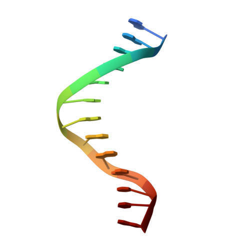
5'-D(*GP*GP*GP*GP*GP*AP*AP*GP*GP*AP *CP*TP*A)-3'
C, E
13N/A
Sequence Annotations
Expand
  • Reference Sequence

Find similar nucleic acids by:  Sequence   |   3D Structure  

Entity ID: 3
MoleculeChains LengthOrganismImage
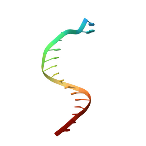
5'-D(*TP*TP*CP*AP*GP*DFTP*AP*GP*TP*CP*CP *TP*TP*CP*CP*CP*CP*C)-3'
D, F
18N/A
Sequence Annotations
Expand
  • Reference Sequence
Small Molecules
Ligands 2 Unique
IDChains Name / Formula / InChI Key2D Diagram3D Interactions
DCT
Query on DCT

Download Ideal Coordinates CCD File 
I [auth A],
N [auth E]
2',3'-DIDEOXYCYTIDINE 5'-TRIPHOSPHATE
C9 H16 N3 O12 P3
ARLKCWCREKRROD-POYBYMJQSA-N
CA
Query on CA

Download Ideal Coordinates CCD File 
G [auth A]
H [auth A]
J [auth A]
K [auth B]
L [auth B]
G [auth A],
H [auth A],
J [auth A],
K [auth B],
L [auth B],
M [auth B]
CALCIUM ION
Ca
BHPQYMZQTOCNFJ-UHFFFAOYSA-N
Experimental Data & Validation

Experimental Data

  • Method: X-RAY DIFFRACTION
  • Resolution: 2.80 Å
  • R-Value Free: 0.250 
  • R-Value Work: 0.211 
  • R-Value Observed: 0.211 
  • Space Group: P 1 21 1
Unit Cell:
Length ( Å )Angle ( ˚ )
a = 52.139α = 90
b = 101.875β = 94.92
c = 111.062γ = 90
Software Package:
Software NamePurpose
CNSrefinement
XDSdata reduction
XDSdata scaling
PHASERphasing

Structure Validation

View Full Validation Report


Ligand Structure Quality Assessment 


Entry History 

Deposition Data

Revision History  (Full details and data files)

  • Version 1.0: 2007-10-30
    Type: Initial release
  • Version 1.1: 2014-04-09
    Changes: Atomic model, Database references, Derived calculations, Non-polymer description, Other, Structure summary, Version format compliance
  • Version 1.2: 2023-12-13
    Changes: Data collection, Database references, Derived calculations, Other, Refinement description