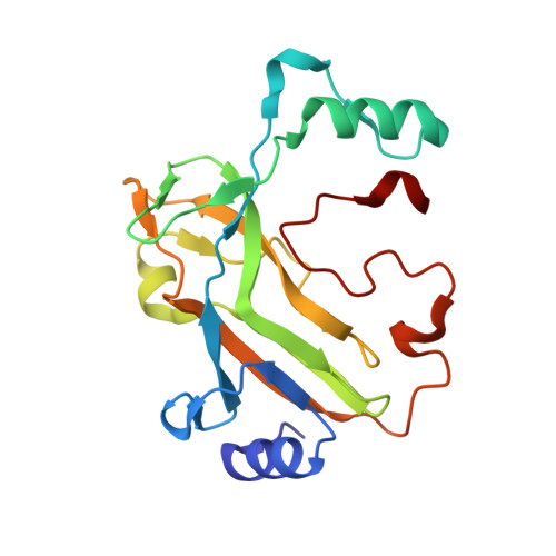
2V9X

E138D variant of Escherichia coli dCTP deaminase in complex with dUTP


Experimental Data Snapshot

  • Method: X-RAY DIFFRACTION
  • Resolution: 2.20 Å
  • R-Value Free: 0.239 
  • R-Value Work: 0.203 
  • R-Value Observed: 0.205 

Starting Model: experimental
View more details

wwPDB Validation   3D Report Full Report


Ligand Structure Quality Assessment 


This is version 1.4 of the entry. See complete history


Literature

Mutational Analysis of the Nucleotide Binding Site of Escherichia Coli Dctp Deaminase.

Thymark, M.Johansson, E.Larsen, S.Willemoes, M.

(2008) Arch Biochem Biophys 470: 20

  • DOI: https://doi.org/10.1016/j.abb.2007.10.013
  • Primary Citation of Related Structures:  
    2V9X

  • PubMed Abstract: 

    In Escherichia coli and Salmonella typhimurium about 80% of the dUMP used for dTMP synthesis is derived from deamination of dCTP. The dCTP deaminase produces dUTP that subsequently is hydrolyzed by dUTPase to dUMP and diphosphate. The dCTP deaminase is regulated by dTTP that inhibits the enzyme by binding to the active site and induces an inactive conformation of the trimeric enzyme. We have analyzed the role of residues previously suggested to play a role in catalysis. The mutant enzymes R115Q, S111C, S111T and E138D were all purified and analyzed for activity. Only S111T and E138D displayed detectable activity with a 30- and 140-fold reduction in k(cat), respectively. Furthermore, S111T and E138D both showed altered dTTP inhibition compared to wild-type enzyme. S111T was almost insensitive to the presence of dTTP. With the E138D enzyme the dTTP dependent increase in cooperativity of dCTP saturation was absent, although the dTTP inhibition itself was still cooperative. Modeling of the active site of the S111T enzyme indicated that this enzyme is restricted in forming the inactive dTTP binding conformer due to steric hindrance by the additional methyl group in threonine. The crystal structure of E138D in complex with dUTP showed a hydrogen bonding network in the active site similar to wild-type enzyme. However, changes in the hydrogen bond lengths between the carboxylate and a catalytic water molecule as well as a slightly different orientation of the pyrimidine ring of the bound nucleotide may provide an explanation for the reduced activity.


  • Organizational Affiliation

    Department of Chemistry, University of Copenhagen, Universitetsparken 5, DK-2100 Copenhagen Ø, Denmark.


Macromolecules
Find similar proteins by:  (by identity cutoff)  |  3D Structure
Entity ID: 1
MoleculeChains Sequence LengthOrganismDetailsImage
DEOXYCYTIDINE TRIPHOSPHATE DEAMINASE
A, B, C, D, E
A, B, C, D, E, F, G, H, I, J, K, L
193Escherichia coliMutation(s): 1 
EC: 3.5.4.13
UniProt
Find proteins for P28248 (Escherichia coli (strain K12))
Explore P28248 
Go to UniProtKB:  P28248
Entity Groups  
Sequence Clusters30% Identity50% Identity70% Identity90% Identity95% Identity100% Identity
UniProt GroupP28248
Sequence Annotations
Expand
  • Reference Sequence
Small Molecules
Ligands 3 Unique
IDChains Name / Formula / InChI Key2D Diagram3D Interactions
DUT
Query on DUT

Download Ideal Coordinates CCD File 
AA [auth G]
DA [auth H]
FA [auth I]
HA [auth J]
JA [auth K]
AA [auth G],
DA [auth H],
FA [auth I],
HA [auth J],
JA [auth K],
M [auth A],
MA [auth L],
O [auth B],
Q [auth C],
T [auth D],
W [auth E],
Y [auth F]
DEOXYURIDINE-5'-TRIPHOSPHATE
C9 H15 N2 O14 P3
AHCYMLUZIRLXAA-SHYZEUOFSA-N
SO4
Query on SO4

Download Ideal Coordinates CCD File 
CA [auth G],
LA [auth K],
S [auth C],
V [auth D]
SULFATE ION
O4 S
QAOWNCQODCNURD-UHFFFAOYSA-L
MG
Query on MG

Download Ideal Coordinates CCD File 
BA [auth G]
EA [auth H]
GA [auth I]
IA [auth J]
KA [auth K]
BA [auth G],
EA [auth H],
GA [auth I],
IA [auth J],
KA [auth K],
N [auth A],
NA [auth L],
P [auth B],
R [auth C],
U [auth D],
X [auth E],
Z [auth F]
MAGNESIUM ION
Mg
JLVVSXFLKOJNIY-UHFFFAOYSA-N
Experimental Data & Validation

Experimental Data

  • Method: X-RAY DIFFRACTION
  • Resolution: 2.20 Å
  • R-Value Free: 0.239 
  • R-Value Work: 0.203 
  • R-Value Observed: 0.205 
  • Space Group: P 21 21 21
Unit Cell:
Length ( Å )Angle ( ˚ )
a = 94.187α = 90
b = 71.301β = 90
c = 319.916γ = 90
Software Package:
Software NamePurpose
REFMACrefinement
DENZOdata reduction
SCALEPACKdata scaling
MOLREPphasing

Structure Validation

View Full Validation Report


Ligand Structure Quality Assessment 


Entry History 

Deposition Data

Revision History  (Full details and data files)

  • Version 1.0: 2007-11-20
    Type: Initial release
  • Version 1.1: 2011-05-08
    Changes: Version format compliance
  • Version 1.2: 2011-07-13
    Changes: Version format compliance
  • Version 1.3: 2018-01-17
    Changes: Data collection
  • Version 1.4: 2023-12-13
    Changes: Data collection, Database references, Derived calculations, Other, Refinement description