2V9A | pdb_00002v9a

Structure of Citrate-free Periplasmic Domain of Sensor Histidine Kinase CitA


Experimental Data Snapshot

  • Method: X-RAY DIFFRACTION
  • Resolution: 2.00 Å
  • R-Value Free: 
    0.278 (Depositor) 
  • R-Value Work: 
    0.212 (Depositor), 0.221 (DCC) 

Starting Model: experimental
View more details

wwPDB Validation 3D Report Full Report

Validation slider image for 2V9A

This is version 1.5 of the entry. See complete history

Literature

A Ligand-Induced Switch in the Periplasmic Domain of Sensor Histidine Kinase Cita.

Sevvana, M.Vijayan, V.Zweckstetter, M.Reinelt, S.Madden, D.R.Herbst-Irmer, R.Sheldrick, G.M.Bott, M.Griesinger, C.Becker, S.

(2008) J Mol Biology 377: 512

  • DOI: https://doi.org/10.1016/j.jmb.2008.01.024
  • Primary Citation Related Structures: 
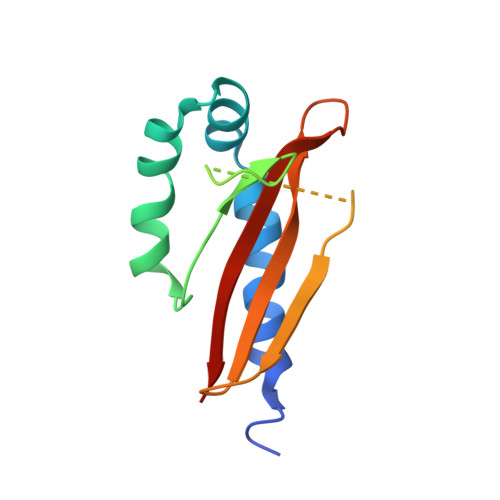
    2J80, 2V9A

  • PubMed Abstract: 

    Sensor histidine kinases of two-component signal-transduction systems are essential for bacteria to adapt to variable environmental conditions. However, despite their prevalence, it is not well understood how extracellular signals such as ligand binding regulate the activity of these sensor kinases. CitA is the sensor histidine kinase in Klebsiella pneumoniae that regulates the transport and anaerobic metabolism of citrate in response to its extracellular concentration. We report here the X-ray structures of the periplasmic sensor domain of CitA in the citrate-free and citrate-bound states. A comparison of the two structures shows that ligand binding causes a considerable contraction of the sensor domain. This contraction may represent the molecular switch that activates transmembrane signaling in the receptor.


  • Organizational Affiliation
    • Department of Structural Chemistry, University of Göttingen, Tammannstrasse 4, 37077 Göttingen, Germany.

Macromolecule Content 

  • Total Structure Weight: 29 kDa 
  • Atom Count: 1,725 
  • Modeled Residue Count: 211 
  • Deposited Residue Count: 266 
  • Unique protein chains: 1

Macromolecules

Find similar proteins by:|  3D Structure
Entity ID: 1
MoleculeChains  Sequence LengthOrganismDetailsImage
SENSOR KINASE CITA
A, B
133Klebsiella pneumoniaeMutation(s): 0 
EC: 2.7.13.3
UniProt
Find proteins for P52687 (Klebsiella pneumoniae)
Explore P52687 
Go to UniProtKB:  P52687
Entity Groups
Sequence Clusters30% Identity50% Identity70% Identity90% Identity95% Identity100% Identity
UniProt GroupP52687
Sequence Annotations
Expand
Reference Sequence

Experimental Data & Validation

Experimental Data

  • Method: X-RAY DIFFRACTION
  • Resolution: 2.00 Å
  • R-Value Free:  0.278 (Depositor) 
  • R-Value Work:  0.212 (Depositor), 0.221 (DCC) 
Space Group: I 4
Unit Cell:
Length ( Å )Angle ( ˚ )
a = 64.163α = 90
b = 64.163β = 90
c = 144.746γ = 90
Software Package:
Software NamePurpose
PHENIXrefinement
DENZOdata reduction
SCALEPACKdata scaling
PHASERphasing
PHENIXrefinement

Structure Validation

View Full Validation Report


Entry History 

Deposition Data

Revision History  (Full details and data files)

  • Version 1.0: 2008-03-25
    Type: Initial release
  • Version 1.1: 2011-05-08
    Changes: Version format compliance
  • Version 1.2: 2011-07-13
    Changes: Version format compliance
  • Version 1.3: 2019-03-06
    Changes: Data collection, Experimental preparation, Other
  • Version 1.4: 2019-05-08
    Changes: Data collection, Experimental preparation
  • Version 1.5: 2023-12-13
    Changes: Data collection, Database references, Other, Refinement description