2V8V | pdb_00002v8v

Crystal structure of mutant R322A of beta-alanine synthase from Saccharomyces kluyveri


Experimental Data Snapshot

  • Method: X-RAY DIFFRACTION
  • Resolution: 2.90 Å
  • R-Value Free: 
    0.246 (Depositor), 0.247 (DCC) 
  • R-Value Work: 
    0.194 (Depositor), 0.198 (DCC) 
  • R-Value Observed: 
    0.196 (Depositor) 

Starting Model: experimental
View more details

wwPDB Validation 3D Report Full Report

Validation slider image for 2V8V

This is version 1.5 of the entry. See complete history

Literature

Crystal Structures of Yeast -Alanine Synthase Complexes Reveal the Mode of Substrate Binding and Large Scale Domain Closure Movements.

Lundgren, S.Andersen, B.Piskur, J.Dobritzsch, D.

(2007) J Biological Chem 282: 36037

  • DOI: https://doi.org/10.1074/jbc.M705517200
  • Primary Citation Related Structures: 
    2V8D, 2V8G, 2V8H, 2V8V

  • PubMed Abstract: 

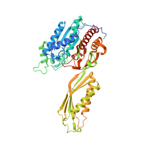
    Beta-alanine synthase is the final enzyme of the reductive pyrimidine catabolic pathway, which is responsible for the breakdown of uracil and thymine in higher organisms. The fold of the homodimeric enzyme from the yeast Saccharomyces kluyveri identifies it as a member of the AcyI/M20 family of metallopeptidases. Its subunit consists of a catalytic domain harboring a di-zinc center and a smaller dimerization domain. The present site-directed mutagenesis studies identify Glu(159) and Arg(322) as crucial for catalysis and His(262) and His(397) as functionally important but not essential. We determined the crystal structures of wild-type beta-alanine synthase in complex with the reaction product beta-alanine, and of the mutant E159A with the substrate N-carbamyl-beta-alanine, revealing the closed state of a dimeric AcyI/M20 metallopeptidase-like enzyme. Subunit closure is achieved by a approximately 30 degrees rigid body domain rotation, which completes the active site by integration of substrate binding residues that belong to the dimerization domain of the same or the partner subunit. Substrate binding is achieved via a salt bridge, a number of hydrogen bonds, and coordination to one of the zinc ions of the di-metal center.


  • Organizational Affiliation
    • Department of Medical Biochemistry and Biophysics, Karolinska Institutet, SE-17177 Stockholm, Sweden.

Macromolecule Content 

  • Total Structure Weight: 208.76 kDa 
  • Atom Count: 13,410 
  • Modeled Residue Count: 1,732 
  • Deposited Residue Count: 1,896 
  • Unique protein chains: 1

Macromolecules

Find similar proteins by:|  3D Structure
Entity ID: 1
MoleculeChains  Sequence LengthOrganismDetailsImage
BETA-ALANINE SYNTHASE
A, B, C, D
474Lachancea kluyveriMutation(s): 1 
EC: 3.5.1.6
UniProt
Find proteins for Q96W94 (Lachancea kluyveri)
Explore Q96W94 
Go to UniProtKB:  Q96W94
Entity Groups
Sequence Clusters30% Identity50% Identity70% Identity90% Identity95% Identity100% Identity
UniProt GroupQ96W94
Sequence Annotations
Expand
Reference Sequence

Small Molecules

Ligands 3 Unique
IDChains Name / Formula / InChI Key2D Diagram3D Interactions
DTT

Query on DTT


Download:Ideal Coordinates CCD File
L [auth C]2,3-DIHYDROXY-1,4-DITHIOBUTANE
C4 H10 O2 S2
VHJLVAABSRFDPM-IMJSIDKUSA-N
URP

Query on URP


Download:Ideal Coordinates CCD File
I [auth B],
M [auth C],
P [auth D]
N-(AMINOCARBONYL)-BETA-ALANINE
C4 H8 N2 O3
JSJWCHRYRHKBBW-UHFFFAOYSA-N
ZN

Query on ZN


Download:Ideal Coordinates CCD File
E [auth A]
F [auth A]
G [auth B]
H [auth B]
J [auth C]
E [auth A],
F [auth A],
G [auth B],
H [auth B],
J [auth C],
K [auth C],
N [auth D],
O [auth D]
ZINC ION
Zn
PTFCDOFLOPIGGS-UHFFFAOYSA-N

Experimental Data & Validation

Experimental Data

  • Method: X-RAY DIFFRACTION
  • Resolution: 2.90 Å
  • R-Value Free:  0.246 (Depositor), 0.247 (DCC) 
  • R-Value Work:  0.194 (Depositor), 0.198 (DCC) 
  • R-Value Observed: 0.196 (Depositor) 
Space Group: P 1
Unit Cell:
Length ( Å )Angle ( ˚ )
a = 77.478α = 67.45
b = 84.903β = 67.81
c = 104.848γ = 62.75
Software Package:
Software NamePurpose
REFMACrefinement
SCALAdata scaling
MOLREPphasing

Structure Validation

View Full Validation Report


Entry History 

Deposition Data

Revision History  (Full details and data files)

  • Version 1.0: 2007-10-02
    Type: Initial release
  • Version 1.1: 2011-07-13
    Changes: Advisory, Refinement description, Version format compliance
  • Version 1.2: 2019-01-30
    Changes: Data collection, Experimental preparation, Other
  • Version 1.3: 2019-02-06
    Changes: Data collection, Experimental preparation
  • Version 1.4: 2023-12-13
    Changes: Data collection, Database references, Derived calculations, Other, Refinement description, Structure summary
  • Version 1.5: 2024-11-13
    Changes: Structure summary