2V5K | pdb_00002v5k

Class II aldolase HpcH - magnesium - oxamate complex


Experimental Data Snapshot

  • Method: X-RAY DIFFRACTION
  • Resolution: 2.20 Å
  • R-Value Free: 
    0.192 (Depositor), 0.184 (DCC) 
  • R-Value Work: 
    0.171 (Depositor), 0.164 (DCC) 
  • R-Value Observed: 
    0.171 (Depositor) 

Starting Model: experimental
View more details

wwPDB Validation 3D Report Full Report

Validation slider image for 2V5K

This is version 1.2 of the entry. See complete history

Literature

Structure and Mechanism of Hpch: A Metal Ion Dependent Class II Aldolase from the Homoprotocatechuate Degradation Pathway of Escherichia Coli.

Rea, D.Fulop, V.Bugg, T.D.H.Roper, D.I.

(2007) J Mol Biology 373: 866

  • DOI: https://doi.org/10.1016/j.jmb.2007.06.048
  • Primary Citation Related Structures: 
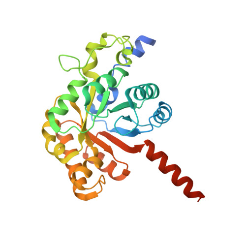
    2V5J, 2V5K

  • PubMed Abstract: 

    Microorganisms are adept at degrading chemically resistant aromatic compounds. One of the longest and most well characterized aromatic catabolic pathways is the 4-hydroxyphenylacetic acid degradation pathway of Escherichia coli. The final step involves the conversion of 4-hydroxy-2-oxo-heptane-1,7-dioate into pyruvate and succinic semialdehyde. This reaction is catalyzed by 4-hydroxy-2-oxo-heptane-1,7-dioate aldolase (HpcH), a member of the divalent metal ion dependent class II aldolase enzymes that have great biosynthetic potential. We have solved the crystal structure of HpcH in the apo form, and with magnesium and the substrate analogue oxamate bound, to 1.6 A and 2.0 A, respectively. Comparison with similar structures of the homologous 2-dehydro-3-deoxygalactarate aldolase, coupled with site-directed mutagenesis data, implicate histidine 45 and arginine 70 as key catalytic residues.


  • Organizational Affiliation
    • Department of Biological Sciences, University of Warwick, Gibbet Hill Road, Coventry, CV4 7AL, UK.

Macromolecule Content 

  • Total Structure Weight: 62.69 kDa 
  • Atom Count: 4,264 
  • Modeled Residue Count: 522 
  • Deposited Residue Count: 574 
  • Unique protein chains: 1

Macromolecules

Find similar proteins by:|  3D Structure
Entity ID: 1
MoleculeChains  Sequence LengthOrganismDetailsImage
2,4-DIHYDROXYHEPT-2-ENE-1,7-DIOIC ACID ALDOLASE
A, B
287Escherichia coli CMutation(s): 0 
EC: 4.1.2 (PDB Primary Data), 4.1.2.52 (UniProt)
UniProt
Find proteins for Q47098 (Escherichia coli)
Explore Q47098 
Go to UniProtKB:  Q47098
Entity Groups
Sequence Clusters30% Identity50% Identity70% Identity90% Identity95% Identity100% Identity
UniProt GroupQ47098
Sequence Annotations
Expand
Reference Sequence

Experimental Data & Validation

Experimental Data

  • Method: X-RAY DIFFRACTION
  • Resolution: 2.20 Å
  • R-Value Free:  0.192 (Depositor), 0.184 (DCC) 
  • R-Value Work:  0.171 (Depositor), 0.164 (DCC) 
  • R-Value Observed: 0.171 (Depositor) 
Space Group: P 21 3
Unit Cell:
Length ( Å )Angle ( ˚ )
a = 152.2α = 90
b = 152.2β = 90
c = 152.2γ = 90
Software Package:
Software NamePurpose
REFMACrefinement
DENZOdata reduction
SCALEPACKdata scaling
PHASERphasing

Structure Validation

View Full Validation Report


Entry History 

Deposition Data

Revision History  (Full details and data files)

  • Version 1.0: 2007-10-02
    Type: Initial release
  • Version 1.1: 2011-07-13
    Changes: Advisory, Version format compliance
  • Version 1.2: 2023-12-13
    Changes: Data collection, Database references, Derived calculations, Other, Refinement description