2V4Z

The crystal structure of the human G-protein subunit alpha (GNAI3) in complex with an engineered regulator of G-protein signaling type 2 domain (RGS2)


Experimental Data Snapshot

  • Method: X-RAY DIFFRACTION
  • Resolution: 2.80 Å
  • R-Value Free: 0.254 
  • R-Value Work: 0.208 
  • R-Value Observed: 0.210 

wwPDB Validation   3D Report Full Report


Ligand Structure Quality Assessment 


This is version 1.3 of the entry. See complete history


Literature

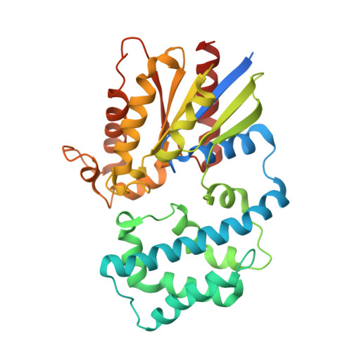
Structural Determinants of G-Protein Alpha Subunit Selectivity by Regulator of G-Protein Signaling 2(Rgs2).

Kimple, A.J.Soundararajan, M.Hutsell, S.Q.Roos, A.K.Urban, D.J.Setola, V.Temple, B.R.Roth, B.L.Knapp, S.Willard, F.S.Siderovski, D.P.

(2009) J Biol Chem 284: 19402

  • DOI: https://doi.org/10.1074/jbc.M109.024711
  • Primary Citation of Related Structures:  
    2V4Z

  • PubMed Abstract: 

    "Regulator of G-protein signaling" (RGS) proteins facilitate the termination of G protein-coupled receptor (GPCR) signaling via their ability to increase the intrinsic GTP hydrolysis rate of Galpha subunits (known as GTPase-accelerating protein or "GAP" activity). RGS2 is unique in its in vitro potency and selectivity as a GAP for Galpha(q) subunits. As many vasoconstrictive hormones signal via G(q) heterotrimer-coupled receptors, it is perhaps not surprising that RGS2-deficient mice exhibit constitutive hypertension. However, to date the particular structural features within RGS2 determining its selectivity for Galpha(q) over Galpha(i/o) substrates have not been completely characterized. Here, we examine a trio of point mutations to RGS2 that elicits Galpha(i)-directed binding and GAP activities without perturbing its association with Galpha(q). Using x-ray crystallography, we determined a model of the triple mutant RGS2 in complex with a transition state mimetic form of Galpha(i) at 2.8-A resolution. Structural comparison with unliganded, wild type RGS2 and of other RGS domain/Galpha complexes highlighted the roles of these residues in wild type RGS2 that weaken Galpha(i) subunit association. Moreover, these three amino acids are seen to be evolutionarily conserved among organisms with modern cardiovascular systems, suggesting that RGS2 arose from the R4-subfamily of RGS proteins to have specialized activity as a potent and selective Galpha(q) GAP that modulates cardiovascular function.


  • Organizational Affiliation

    Departments of Pharmacology, University of North Carolina, Chapel Hill, North Carolina 27599, USA.


Macromolecules
Find similar proteins by:  (by identity cutoff)  |  3D Structure
Entity ID: 1
MoleculeChains Sequence LengthOrganismDetailsImage
GUANINE NUCLEOTIDE-BINDING PROTEIN G(K) SUBUNIT ALPHA350Homo sapiensMutation(s): 0 
UniProt & NIH Common Fund Data Resources
Find proteins for P08754 (Homo sapiens)
Explore P08754 
Go to UniProtKB:  P08754
PHAROS:  P08754
GTEx:  ENSG00000065135 
Entity Groups  
Sequence Clusters30% Identity50% Identity70% Identity90% Identity95% Identity100% Identity
UniProt GroupP08754
Sequence Annotations
Expand
  • Reference Sequence
Find similar proteins by:  (by identity cutoff)  |  3D Structure
Entity ID: 2
MoleculeChains Sequence LengthOrganismDetailsImage
REGULATOR OF G-PROTEIN SIGNALING 2142Homo sapiensMutation(s): 3 
UniProt & NIH Common Fund Data Resources
Find proteins for P41220 (Homo sapiens)
Explore P41220 
Go to UniProtKB:  P41220
PHAROS:  P41220
GTEx:  ENSG00000116741 
Entity Groups  
Sequence Clusters30% Identity50% Identity70% Identity90% Identity95% Identity100% Identity
UniProt GroupP41220
Sequence Annotations
Expand
  • Reference Sequence
Experimental Data & Validation

Experimental Data

  • Method: X-RAY DIFFRACTION
  • Resolution: 2.80 Å
  • R-Value Free: 0.254 
  • R-Value Work: 0.208 
  • R-Value Observed: 0.210 
  • Space Group: P 32 2 1
Unit Cell:
Length ( Å )Angle ( ˚ )
a = 114.539α = 90
b = 114.539β = 90
c = 99.33γ = 120
Software Package:
Software NamePurpose
REFMACrefinement
MOSFLMdata reduction
SCALAdata scaling
PHASERphasing

Structure Validation

View Full Validation Report


Ligand Structure Quality Assessment 


Entry History 

Deposition Data

Revision History  (Full details and data files)

  • Version 1.0: 2008-11-04
    Type: Initial release
  • Version 1.1: 2011-07-13
    Changes: Advisory, Version format compliance
  • Version 1.2: 2018-04-04
    Changes: Data collection
  • Version 1.3: 2023-12-13
    Changes: Data collection, Database references, Derived calculations, Other, Refinement description