2UZS | pdb_00002uzs

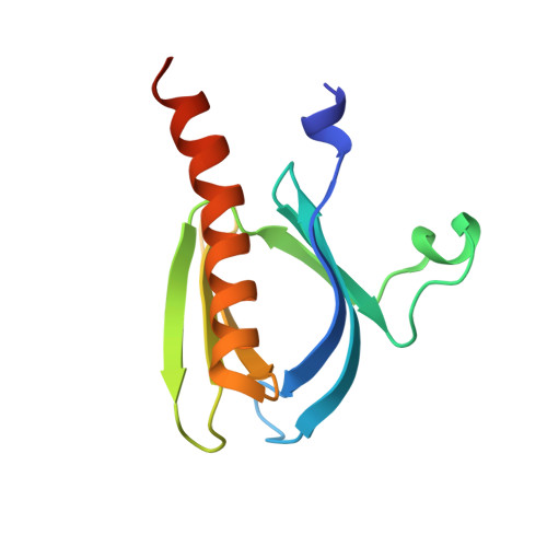
A transforming mutation in the pleckstrin homology domain of AKT1 in cancer (AKT1-PH_E17K)


Experimental Data Snapshot

  • Method: X-RAY DIFFRACTION
  • Resolution: 2.46 Å
  • R-Value Free: 
    0.286 (Depositor) 
  • R-Value Work: 
    0.207 (Depositor) 
  • R-Value Observed: 
    0.211 (Depositor) 

Starting Model: experimental
View more details

wwPDB Validation 3D Report Full Report

Validation slider image for 2UZS

Ligand Structure Quality Assessment 


This is version 1.6 of the entry. See complete history. 

Literature

A transforming mutation in the pleckstrin homology domain of AKT1 in cancer.

Carpten, J.D., Faber, A.L., Horn, C., Donoho, G.P., Briggs, S.L., Robbins, C.M., Hostetter, G., Boguslawski, S., Moses, T.Y., Savage, S., Uhlik, M., Lin, A., Du, J., Qian, Y.W., Zeckner, D.J., Tucker-Kellogg, G., Touchman, J., Patel, K., Mousses, S., Bittner, M., Schevitz, R., Lai, M.H., Blanchard, K.L., Thomas, J.E.

(2007) Nature 448: 439-444

  • DOI: https://doi.org/10.1038/nature05933
  • Primary Citation Related Structures: 
    2UZR, 2UZS

  • PubMed Abstract: 

    Although AKT1 (v-akt murine thymoma viral oncogene homologue 1) kinase is a central member of possibly the most frequently activated proliferation and survival pathway in cancer, mutation of AKT1 has not been widely reported. Here we report the identification of a somatic mutation in human breast, colorectal and ovarian cancers that results in a glutamic acid to lysine substitution at amino acid 17 (E17K) in the lipid-binding pocket of AKT1. Lys 17 alters the electrostatic interactions of the pocket and forms new hydrogen bonds with a phosphoinositide ligand. This mutation activates AKT1 by means of pathological localization to the plasma membrane, stimulates downstream signalling, transforms cells and induces leukaemia in mice. This mechanism indicates a direct role of AKT1 in human cancer, and adds to the known genetic alterations that promote oncogenesis through the phosphatidylinositol-3-OH kinase/AKT pathway. Furthermore, the E17K substitution decreases the sensitivity to an allosteric kinase inhibitor, so this mutation may have important clinical utility for AKT drug development.


  • Organizational Affiliation: 
    • Division of Integrated Cancer Genomics, Translational Genomics Research Institute, 445 N. Fifth Street, Phoenix, Arizona 85004, USA.

Macromolecule Content 

  • Total Structure Weight: 15.3 kDa 
  • Atom Count: 1,053 
  • Modeled Residue Count: 119 
  • Deposited Residue Count: 125 
  • Unique protein chains: 1

Macromolecules

Find similar proteins by:|  3D Structure
Entity ID: 1
MoleculeChains  Sequence LengthOrganismDetailsImage
RAC-alpha serine/threonine-protein kinase125Homo sapiensMutation(s): 1 
Gene Names: AKT1, PKB, RAC
EC: 2.7.11.1
UniProt & NIH Common Fund Data Resources
Find proteins for P31749 (Homo sapiens)
Explore P31749 
Go to UniProtKB:  P31749
PHAROS:  P31749
GTEx:  ENSG00000142208 
Entity Groups
Sequence Clusters30% Identity50% Identity70% Identity90% Identity95% Identity100% Identity
UniProt GroupP31749
Sequence Annotations
Expand
Reference Sequence

Small Molecules

Ligands 1 Unique
IDChains Name / Formula / InChI Key2D Diagram3D Interactions
4IP

Query on 4IP


Download:Ideal Coordinates CCD File
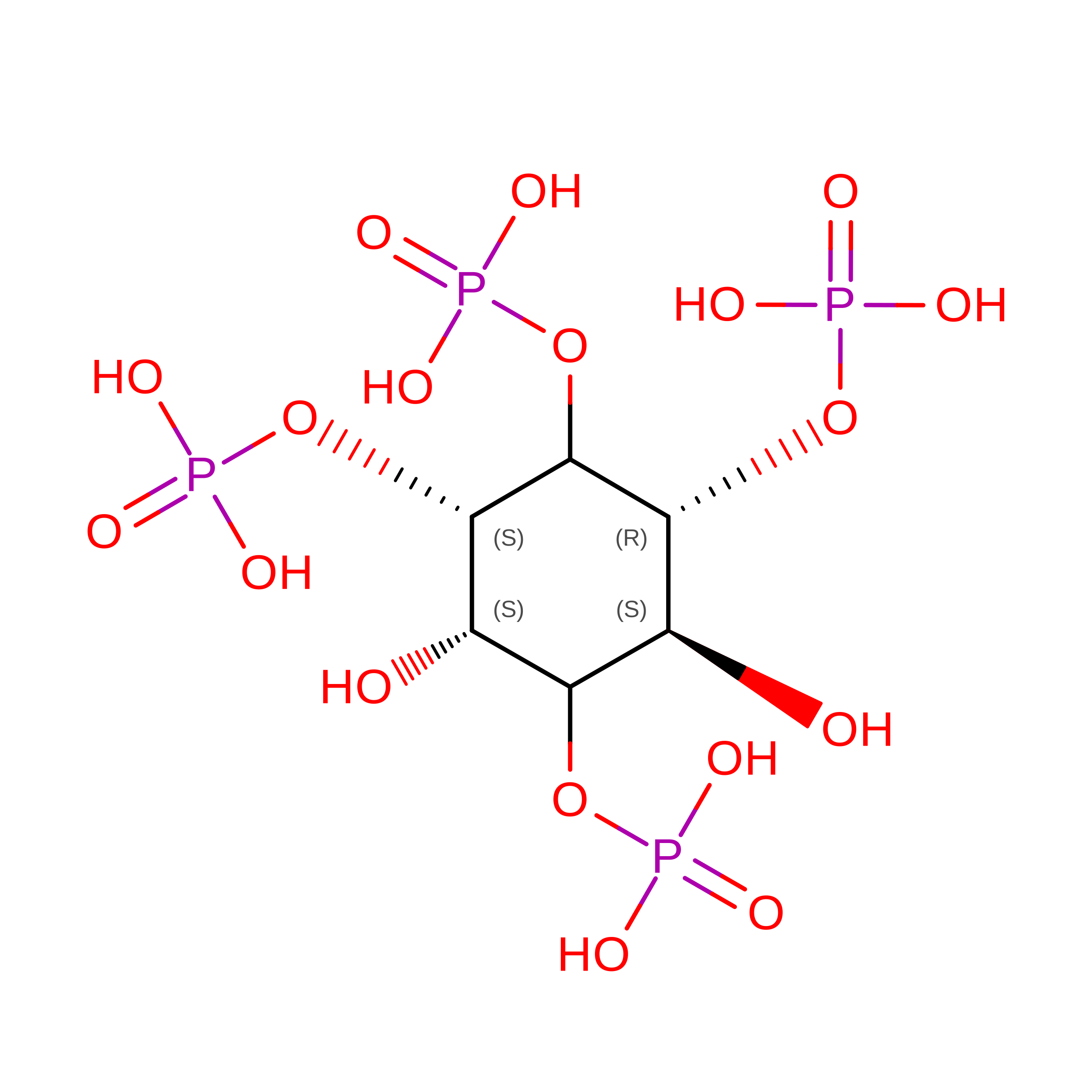
B [auth A]INOSITOL-(1,3,4,5)-TETRAKISPHOSPHATE
C6 H16 O18 P4
CIPFCGZLFXVXBG-CNWJWELYSA-N

Experimental Data & Validation

Experimental Data

  • Method: X-RAY DIFFRACTION
  • Resolution: 2.46 Å
  • R-Value Free:  0.286 (Depositor) 
  • R-Value Work:  0.207 (Depositor) 
  • R-Value Observed: 0.211 (Depositor) 
Space Group: C 1 2 1
Unit Cell:
Length ( Å )Angle ( ˚ )
a = 79.299α = 90
b = 32.711β = 116.67
c = 41.983γ = 90
Software Package:
Software NamePurpose
REFMACrefinement
DENZOdata reduction
SCALEPACKdata scaling
AMoREphasing

Structure Validation

View Full Validation Report


Ligand Structure Quality Assessment 


Entry History 

Revision History  (Full details and data files)

  • Version 1.0: 2007-07-17
    Type: Initial release
  • Version 1.1: 2011-05-08
    Changes: Version format compliance
  • Version 1.2: 2011-07-13
    Changes: Version format compliance
  • Version 1.3: 2018-10-24
    Changes: Data collection, Database references, Source and taxonomy, Structure summary
  • Version 1.4: 2019-05-08
    Changes: Data collection, Derived calculations, Experimental preparation
  • Version 1.5: 2023-12-13
    Changes: Data collection, Database references, Other, Refinement description
  • Version 1.6: 2025-10-01
    Changes: Advisory, Derived calculations, Structure summary