2UY1 | pdb_00002uy1

CRYSTAL STRUCTURE OF CSTF-77


Experimental Data Snapshot

  • Method: X-RAY DIFFRACTION
  • Resolution: 2.00 Å
  • R-Value Free: 
    0.280 (Depositor), 0.221 (DCC) 
  • R-Value Work: 
    0.225 (Depositor), 0.227 (DCC) 
  • R-Value Observed: 
    0.227 (Depositor) 

wwPDB Validation 3D Report Full Report

Validation slider image for 2UY1

This is version 1.3 of the entry. See complete history

Literature

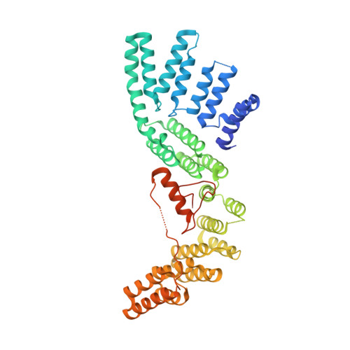
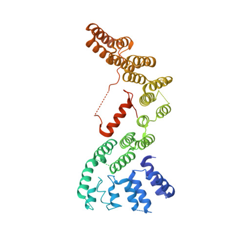
The Structure of the Cstf-77 Homodimer Provides Insights Into Cstf Assembly.

Legrand, P.Pinaud, N.Minvielle-Sebastia, L.Fribourg, S.

(2007) Nucleic Acids Res 35: 4515

  • DOI: https://doi.org/10.1093/nar/gkm458
  • Primary Citation Related Structures: 
    2UY1

  • PubMed Abstract: 

    The cleavage stimulation factor (CstF) is essential for the first step of poly(A) tail formation at the 3' ends of mRNAs. This heterotrimeric complex is built around the 77-kDa protein bridging both CstF-64 and CstF-50 subunits. We have solved the crystal structure of the 77-kDa protein from Encephalitozoon cuniculi at a resolution of 2 A. The structure folds around 11 Half-a-TPR repeats defining two domains. The crystal structure reveals a tight homodimer exposing phylogenetically conserved areas for interaction with protein partners. Mapping experiments identify the C-terminal region of Rna14p, the yeast counterpart of CstF-77, as the docking domain for Rna15p, the yeast CstF-64 homologue.


  • Organizational Affiliation
    • Institut Européen de Chimie et Biologie, INSERM U869, 2 rue Robert Escarpit Pessac, F-33607, Université Victor Segalen, Bordeaux 2.

Macromolecule Content 

  • Total Structure Weight: 116.45 kDa 
  • Atom Count: 7,223 
  • Modeled Residue Count: 822 
  • Deposited Residue Count: 986 
  • Unique protein chains: 2

Macromolecules

Find similar proteins by:|  3D Structure
Entity ID: 1
MoleculeChains  Sequence LengthOrganismDetailsImage
CLEAVAGE STIMULATION FACTOR 77493Encephalitozoon cuniculiMutation(s): 0 
UniProt
Find proteins for Q8SWC5 (Encephalitozoon cuniculi (strain GB-M1))
Explore Q8SWC5 
Go to UniProtKB:  Q8SWC5
Entity Groups
Sequence Clusters30% Identity50% Identity70% Identity90% Identity95% Identity100% Identity
UniProt GroupQ8SWC5
Sequence Annotations
Expand
Reference Sequence
Find similar proteins by:|  3D Structure
Entity ID: 2
MoleculeChains  Sequence LengthOrganismDetailsImage
CLEAVAGE STIMULATION FACTOR 77493Encephalitozoon cuniculiMutation(s): 0 
UniProt
Find proteins for Q8SWC5 (Encephalitozoon cuniculi (strain GB-M1))
Explore Q8SWC5 
Go to UniProtKB:  Q8SWC5
Entity Groups
Sequence Clusters30% Identity50% Identity70% Identity90% Identity95% Identity100% Identity
UniProt GroupQ8SWC5
Sequence Annotations
Expand
Reference Sequence

Experimental Data & Validation

Experimental Data

  • Method: X-RAY DIFFRACTION
  • Resolution: 2.00 Å
  • R-Value Free:  0.280 (Depositor), 0.221 (DCC) 
  • R-Value Work:  0.225 (Depositor), 0.227 (DCC) 
  • R-Value Observed: 0.227 (Depositor) 
Space Group: P 1 21 1
Unit Cell:
Length ( Å )Angle ( ˚ )
a = 44.44α = 90
b = 148.28β = 92.15
c = 91.44γ = 90
Software Package:
Software NamePurpose
REFMACrefinement
XDSdata reduction
XSCALEdata scaling

Structure Validation

View Full Validation Report


Entry History 

Deposition Data

Revision History  (Full details and data files)

  • Version 1.0: 2007-07-03
    Type: Initial release
  • Version 1.1: 2011-05-08
    Changes: Version format compliance
  • Version 1.2: 2011-07-13
    Changes: Version format compliance
  • Version 1.3: 2024-05-08
    Changes: Data collection, Database references, Other