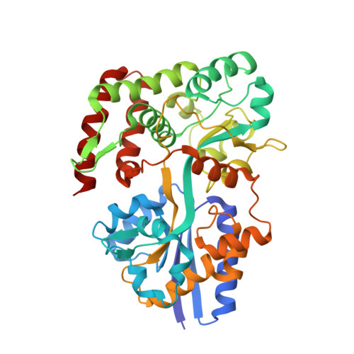
Specific Recognition of Saturated and 4,5-Unsaturated Hexuronate Sugars by a Periplasmic Binding Protein Involved in Pectin Catabolism.
Abbott, D.W., Boraston, A.B.(2007) J Mol Biology 369: 759
- PubMed: 17451747
- DOI: https://doi.org/10.1016/j.jmb.2007.03.045
- Primary Citation of Related Structures:
2UVG, 2UVH, 2UVI, 2UVJ - PubMed Abstract:
The process of pectin depolymerization by pectate lyases and glycoside hydrolases produced by pectinolytic organisms, particularly the phytopathogens from the genus Erwinia, is reasonably well understood. Indeed each extracellular and intracellular catabolic stage has been identified using either genetic, bioinformatic or biochemical approaches. Nevertheless, the molecular details of many of these stages remain unknown. In particular, the mechanism and ligand binding profiles for the transport of pectin degradation products between cellular compartments remain entirely uninvestigated. Here we present the structure of TogB, a 45.7 kDa periplasmic binding protein from Yersinia enterocolitica. This protein is a component of the TogMNAB ABC transporter involved in the periplasmic transport of oligogalacturonides. In addition to the unliganded complex (at 2.2 A), we have also determined the structures of TogB in complex with digalacturonic acid (at 2.2 A), trigalacturonic acid (at 1.8 A) and 4,5-unsaturated digalacutronic acid (at 2.3 A). The molecular determinants of oligogalacturonide binding include a novel salt-bridge between the non-reducing sugar uronate group, selectivity for the unsaturated ligand, and the overall sugar configuration. Complementing this are UV difference and isothermal titration calorimetry experiments that highlight the thermodynamic basis of ligand specificity. The ligand binding profiles of the TogMNAB transporter complex nicely complement pectate lyase-mediated pectin degradation, which is a significant component of pectin depolymerization reactions.
- Biochemistry and Microbiology, University of Victoria, PO Box 3055 STN CSC, Victoria, BC, Canada V8W 3P6.
Organizational Affiliation:


