2UUL | pdb_00002uul

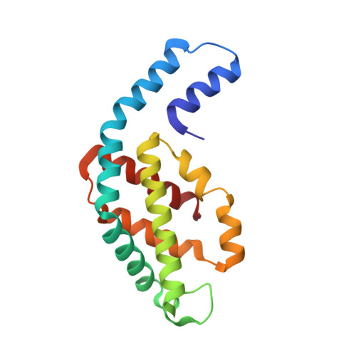
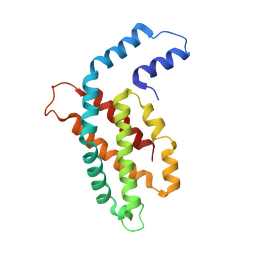
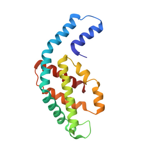
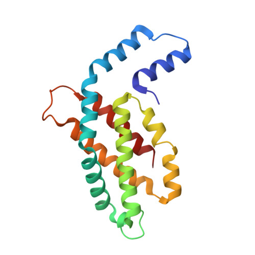
Crystal structure of C-phycocyanin from Phormidium, Lyngbya spp. (Marine) and Spirulina sp. (Fresh water) shows two different ways of energy transfer between two hexamers.


Experimental Data Snapshot

  • Method: X-RAY DIFFRACTION
  • Resolution: 3.10 Å
  • R-Value Free: 
    0.245 (Depositor), 0.241 (DCC) 
  • R-Value Work: 
    0.200 (Depositor), 0.197 (DCC) 
  • R-Value Observed: 
    0.202 (Depositor) 

Starting Model: experimental
View more details

wwPDB Validation 3D Report Full Report

Validation slider image for 2UUL

Ligand Structure Quality Assessment 


This is version 2.0 of the entry. See complete history

Literature

Crystal Structure of C-Phycocyanin from Phormidium, Lyngbya Spp. (Marine) and Spirulina Sp. (Fresh Water) Shows Two Different Ways of Energy Transfer between Twohexamers.

Satyanarayana, L.Patel, A.Mishra, S.Ghosh, P.K.Suresh, C.G.

To be published.

Macromolecule Content 

  • Total Structure Weight: 443.07 kDa 
  • Atom Count: 31,131 
  • Modeled Residue Count: 4,008 
  • Deposited Residue Count: 4,008 
  • Unique protein chains: 5

Macromolecules

Find similar proteins by:|  3D Structure
Entity ID: 1
MoleculeChains  Sequence LengthOrganismDetailsImage
C-PHYCOCYANIN ALPHA CHAIN
A, E, G, I, K
A, E, G, I, K, M, O, Q, S, U, W
162Phormidium sp.Mutation(s): 0 
Find similar proteins by:|  3D Structure
Entity ID: 2
MoleculeChains  Sequence LengthOrganismDetailsImage
C-PHYCOCYANIN BETA CHAIN
B, F, J, L, N
B, F, J, L, N, P, R, T, V, X
172Phormidium sp.Mutation(s): 0 
Find similar proteins by:|  3D Structure
Entity ID: 3
MoleculeChains  Sequence LengthOrganismDetailsImage
C-PHYCOCYANIN ALPHA CHAIN162Phormidium sp.Mutation(s): 0 
Find similar proteins by:|  3D Structure
Entity ID: 4
MoleculeChains  Sequence LengthOrganismDetailsImage
C-PHYCOCYANIN BETA CHAIN172Phormidium sp.Mutation(s): 0 
Find similar proteins by:|  3D Structure
Entity ID: 5
MoleculeChains  Sequence LengthOrganismDetailsImage
C-PHYCOCYANIN BETA CHAIN172Phormidium sp.Mutation(s): 0 

Small Molecules

Ligands 1 Unique
IDChains Name / Formula / InChI Key2D Diagram3D Interactions
CYC

Query on CYC


Download:Ideal Coordinates CCD File
AA [auth B]
AB [auth T]
BA [auth C]
BB [auth T]
CA [auth D]
AA [auth B],
AB [auth T],
BA [auth C],
BB [auth T],
CA [auth D],
CB [auth U],
DA [auth D],
DB [auth V],
EA [auth E],
EB [auth V],
FA [auth F],
FB [auth W],
GA [auth F],
GB [auth X],
HA [auth G],
HB [auth X],
IA [auth H],
JA [auth H],
KA [auth I],
LA [auth J],
MA [auth J],
NA [auth K],
OA [auth L],
PA [auth L],
QA [auth M],
RA [auth N],
SA [auth N],
TA [auth O],
UA [auth P],
VA [auth P],
WA [auth Q],
XA [auth R],
Y [auth A],
YA [auth R],
Z [auth B],
ZA [auth S]
PHYCOCYANOBILIN
C33 H40 N4 O6
VXTXPYZGDQPMHK-GMXXPEQVSA-N

Experimental Data & Validation

Experimental Data

  • Method: X-RAY DIFFRACTION
  • Resolution: 3.10 Å
  • R-Value Free:  0.245 (Depositor), 0.241 (DCC) 
  • R-Value Work:  0.200 (Depositor), 0.197 (DCC) 
  • R-Value Observed: 0.202 (Depositor) 
Space Group: P 1 21 1
Unit Cell:
Length ( Å )Angle ( ˚ )
a = 107.878α = 90
b = 115.783β = 90.24
c = 183.549γ = 90
Software Package:
Software NamePurpose
REFMACrefinement
DENZOdata reduction
SCALEPACKdata scaling
AMoREphasing

Structure Validation

View Full Validation Report


Ligand Structure Quality Assessment 


Entry History 

Deposition Data

Revision History  (Full details and data files)

  • Version 1.0: 2008-05-27
    Type: Initial release
  • Version 1.1: 2011-05-08
    Changes: Version format compliance
  • Version 1.2: 2011-07-13
    Changes: Version format compliance
  • Version 1.3: 2023-12-13
    Changes: Data collection, Database references, Derived calculations, Other, Refinement description
  • Version 1.4: 2024-11-06
    Changes: Structure summary
  • Version 2.0: 2025-12-10
    Changes: Atomic model, Data collection, Derived calculations, Non-polymer description, Structure summary