2SEB | pdb_00002seb

X-RAY CRYSTAL STRUCTURE OF HLA-DR4 COMPLEXED WITH A PEPTIDE FROM HUMAN COLLAGEN II


Experimental Data Snapshot

  • Method: X-RAY DIFFRACTION
  • Resolution: 2.50 Å
  • R-Value Free: 
    0.280 (Depositor) 
  • R-Value Work: 
    0.229 (Depositor), 0.240 (DCC) 
  • R-Value Observed: 
    0.229 (Depositor) 

Starting Model: experimental
View more details

wwPDB Validation   3D Report Full Report


Ligand Structure Quality Assessment 

Created with Raphaël 2.3.0Worse 01 BetterLigand structure goodness of fit to experimental dataBest fitted NAGClick on this verticalbar to view details

This is version 1.5 of the entry. See complete history


Literature

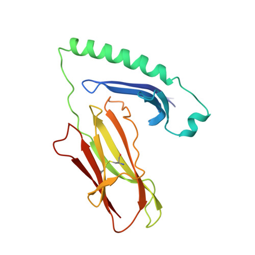
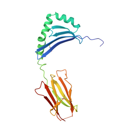
X-ray crystal structure of HLA-DR4 (DRA*0101, DRB1*0401) complexed with a peptide from human collagen II.

Dessen, A.Lawrence, C.M.Cupo, S.Zaller, D.M.Wiley, D.C.

(1997) Immunity 7: 473-481

  • DOI: https://doi.org/10.1016/s1074-7613(00)80369-6
  • Primary Citation of Related Structures:  
    2SEB

  • PubMed Abstract: 

    Genetic predisposition to rheumatoid arthritis (RA) is linked to the MHC class II allele HLA-DR4. The charge of the amino acid at DRbeta71 in the peptide-binding site appears to be critical in discriminating DR molecules linked to increased disease susceptibility. We have determined the 2.5 A x-ray structure of the DR4 molecule with the strongest linkage to RA (DRB1*0401) complexed with a human collagen II peptide. Details of a predicted salt bridge between lysine DRbeta71 and aspartic acid at the P4 peptide position suggest how it may participate in both antigen binding and TCR activation. A model is proposed for the DR4 recognition of collagen II (261-273), an antigen immunodominant in human-transgenic mouse models of RA.


  • Organizational Affiliation

    Laboratory of Molecular Medicine, The Children's Hospital, Boston, Massachusetts 02115, USA. dessen@xtal22.tch.harvard.edu


Macromolecules
Find similar proteins by:  (by identity cutoff)  |  3D Structure
Entity ID: 1
MoleculeChains Sequence LengthOrganismDetailsImage
HLA CLASS II HISTOCOMPATIBILITY ANTIGEN181Homo sapiensMutation(s): 0 
UniProt & NIH Common Fund Data Resources
Find proteins for P01903 (Homo sapiens)
Explore P01903 
Go to UniProtKB:  P01903
PHAROS:  P01903
GTEx:  ENSG00000204287 
Entity Groups  
Sequence Clusters30% Identity50% Identity70% Identity90% Identity95% Identity100% Identity
UniProt GroupP01903
Glycosylation
Glycosylation Sites: 1Go to GlyGen: P01903-1
Sequence Annotations
Expand
  • Reference Sequence
Find similar proteins by:  (by identity cutoff)  |  3D Structure
Entity ID: 2
MoleculeChains Sequence LengthOrganismDetailsImage
HLA CLASS II HISTOCOMPATIBILITY ANTIGEN192Homo sapiensMutation(s): 0 
UniProt & NIH Common Fund Data Resources
Find proteins for P01911 (Homo sapiens)
Explore P01911 
Go to UniProtKB:  P01911
PHAROS:  P01911
GTEx:  ENSG00000196126 
Entity Groups  
Sequence Clusters30% Identity50% Identity70% Identity90% Identity95% Identity100% Identity
UniProt GroupP01911
Sequence Annotations
Expand
  • Reference Sequence
Find similar proteins by:  (by identity cutoff)  |  3D Structure
Entity ID: 3
MoleculeChains Sequence LengthOrganismDetailsImage
ENTEROTOXIN TYPE BC [auth D]239Staphylococcus aureusMutation(s): 0 
UniProt
Find proteins for P01552 (Staphylococcus aureus)
Explore P01552 
Go to UniProtKB:  P01552
Entity Groups  
Sequence Clusters30% Identity50% Identity70% Identity90% Identity95% Identity100% Identity
UniProt GroupP01552
Sequence Annotations
Expand
  • Reference Sequence

Find similar proteins by:  Sequence   |   3D Structure  

Entity ID: 4
MoleculeChains Sequence LengthOrganismDetailsImage
PEPTIDE FROM COLLAGEN IID [auth E]12Homo sapiensMutation(s): 0 
UniProt & NIH Common Fund Data Resources
Find proteins for P02458 (Homo sapiens)
Explore P02458 
Go to UniProtKB:  P02458
PHAROS:  P02458
GTEx:  ENSG00000139219 
Entity Groups  
Sequence Clusters30% Identity50% Identity70% Identity90% Identity95% Identity100% Identity
UniProt GroupP02458
Sequence Annotations
Expand
  • Reference Sequence
Small Molecules
Ligands 1 Unique
IDChains Name / Formula / InChI Key2D Diagram3D Interactions
NAG
Query on NAG

Download Ideal Coordinates CCD File 
E [auth A]2-acetamido-2-deoxy-beta-D-glucopyranose
C8 H15 N O6
OVRNDRQMDRJTHS-FMDGEEDCSA-N
Experimental Data & Validation

Experimental Data

  • Method: X-RAY DIFFRACTION
  • Resolution: 2.50 Å
  • R-Value Free:  0.280 (Depositor) 
  • R-Value Work:  0.229 (Depositor), 0.240 (DCC) 
  • R-Value Observed: 0.229 (Depositor) 
Space Group: P 21 21 21
Unit Cell:
Length ( Å )Angle ( ˚ )
a = 78.53α = 90
b = 100.28β = 90
c = 100.87γ = 90
Software Package:
Software NamePurpose
DENZOdata reduction
SCALEPACKdata scaling
X-PLORmodel building
X-PLORrefinement
X-PLORphasing

Structure Validation

View Full Validation Report


Ligand Structure Quality Assessment 

Created with Raphaël 2.3.0Worse 01 BetterLigand structure goodness of fit to experimental dataBest fitted NAGClick on this verticalbar to view details

Entry History 

Deposition Data

Revision History  (Full details and data files)

  • Version 1.0: 1998-01-28
    Type: Initial release
  • Version 1.1: 2008-03-25
    Changes: Version format compliance
  • Version 1.2: 2011-07-13
    Changes: Non-polymer description, Version format compliance
  • Version 1.3: 2020-07-29
    Type: Remediation
    Reason: Carbohydrate remediation
    Changes: Advisory, Data collection, Database references, Derived calculations, Structure summary
  • Version 1.4: 2023-08-09
    Changes: Advisory, Database references, Refinement description, Structure summary
  • Version 1.5: 2024-10-23
    Changes: Data collection, Structure summary