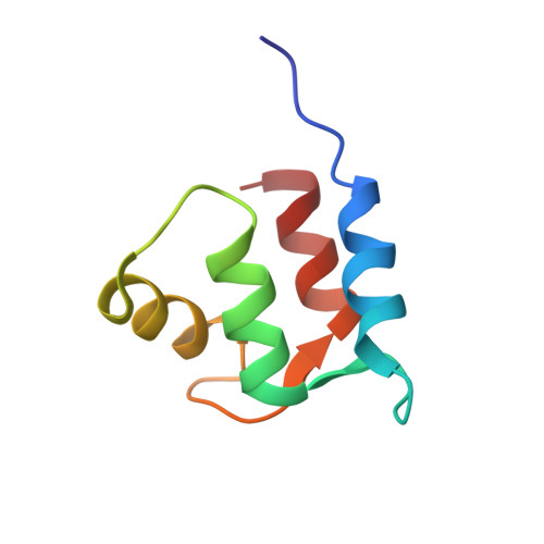
Solution structure and fluctuation of the Mg(2+)-bound form of calmodulin C-terminal domain
Ohashi, W., Hirota, H., Yamazaki, T.(2011) Protein Sci 20: 690-701
- PubMed: 21312310 Search on PubMedSearch on PubMed Central
- DOI: https://doi.org/10.1002/pro.598
- Primary Citation Related Structures:
2RRT - PubMed Abstract:
Calmodulin (CaM) is a Ca(2+)-binding protein that functions as a ubiquitous Ca(2+)-signaling molecule, through conformational changes from the "closed" apo conformation to the "open" Ca(2+)-bound conformation. Mg(2+) also binds to CaM and stabilizes its folded structure, but the NMR signals are broadened by slow conformational fluctuations. Using the E104D/E140D mutant, designed to decrease the signal broadening in the presence of Mg(2+) with minimal perturbations of the overall structure, the solution structure of the Mg(2+)-bound form of the CaM C-terminal domain was determined by multidimensional NMR spectroscopy. The Mg(2+)-induced conformational change mainly occurred in EF hand IV, while EF-hand III retained the apo structure. The helix G and helix H sides of the binding sequence undergo conformational changes needed for the Mg(2+) coordination, and thus the helices tilt slightly. The aromatic rings on helix H move to form a new cluster of aromatic rings in the hydrophobic core. Although helix G tilts slightly to the open orientation, the closed conformation is maintained. The fact that the Mg(2+)-induced conformational changes in EF-hand IV and the hydrophobic core are also seen upon Ca(2+) binding suggests that the Ca(2+)-induced conformational changes can be divided into two categories, those specific to Ca(2+) and those common to Ca(2+) and Mg(2+).
- Genomic Sciences Center, RIKEN, 1-7-22, Suehiro, Tsurumi, Yokohama 230-0045, Japan.
Organizational Affiliation: