2RNM | pdb_00002rnm

Structure of The HET-s(218-289) prion in its amyloid form obtained by solid-state NMR


Experimental Data Snapshot

  • Method: SOLUTION NMR
  • Conformers Calculated: 200 
  • Conformers Submitted: 20 
  • Selection Criteria: structures with the lowest energy 

wwPDB Validation 3D Report Full Report

Validation slider image for 2RNM

This is version 1.4 of the entry. See complete history

Literature

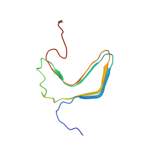
Amyloid fibrils of the HET-s(218-289) prion form a beta solenoid with a triangular hydrophobic core

Wasmer, C.Lange, A.Van Melckebeke, H.Siemer, A.B.Riek, R.Meier, B.H.

(2008) Science 319: 1523-1526

  • DOI: https://doi.org/10.1126/science.1151839
  • Primary Citation Related Structures: 
    2RNM

  • PubMed Abstract: 

    Prion and nonprion forms of proteins are believed to differ solely in their three-dimensional structure, which is therefore of paramount importance for the prion function. However, no atomic-resolution structure of the fibrillar state that is likely infectious has been reported to date. We present a structural model based on solid-state nuclear magnetic resonance restraints for amyloid fibrils from the prion-forming domain (residues 218 to 289) of the HET-s protein from the filamentous fungus Podospora anserina. On the basis of 134 intra- and intermolecular experimental distance restraints, we find that HET-s(218-289) forms a left-handed beta solenoid, with each molecule forming two helical windings, a compact hydrophobic core, at least 23 hydrogen bonds, three salt bridges, and two asparagine ladders. The structure is likely to have broad implications for understanding the infectious amyloid state.


  • Organizational Affiliation
    • Physical Chemistry, ETH Zurich, 8093 Zurich, Switzerland.

Macromolecule Content 

  • Total Structure Weight: 43.34 kDa 
  • Atom Count: 3,045 
  • Modeled Residue Count: 395 
  • Deposited Residue Count: 395 
  • Unique protein chains: 1

Macromolecules

Find similar proteins by:|  3D Structure
Entity ID: 1
MoleculeChains  Sequence LengthOrganismDetailsImage
Small s protein
A, B, C, D, E
79Podospora anserinaMutation(s): 0 
UniProt
Find proteins for Q03689 (Podospora anserina)
Explore Q03689 
Go to UniProtKB:  Q03689
Entity Groups
Sequence Clusters30% Identity50% Identity70% Identity90% Identity95% Identity100% Identity
UniProt GroupQ03689
Sequence Annotations
Expand
Reference Sequence

Experimental Data & Validation

Experimental Data

  • Method: SOLUTION NMR
  • Conformers Calculated: 200 
  • Conformers Submitted: 20 
  • Selection Criteria: structures with the lowest energy 

Structure Validation

View Full Validation Report


Entry History 

Deposition Data

Revision History  (Full details and data files)

  • Version 1.0: 2008-04-01
    Type: Initial release
  • Version 1.1: 2009-02-24
    Changes: Version format compliance
  • Version 1.2: 2015-06-17
    Changes: Version format compliance
  • Version 1.3: 2015-07-08
    Changes: Other
  • Version 1.4: 2024-05-01
    Changes: Data collection, Database references