2RLT | pdb_00002rlt

phosphorylated CPI-17 (22-120)


Experimental Data Snapshot

  • Method: SOLUTION NMR
  • Conformers Calculated: 100 
  • Conformers Submitted: 
  • Selection Criteria: structures with the lowest energy 

wwPDB Validation 3D Report Full Report

Validation slider image for 2RLT

This is version 1.3 of the entry. See complete history

Literature

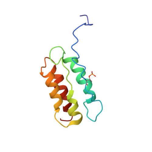
Phosphorylation-induced conformational switching of CPI-17 produces a potent myosin phosphatase inhibitor.

Eto, M.Kitazawa, T.Matsuzawa, F.Aikawa, S.Kirkbride, J.A.Isozumi, N.Nishimura, Y.Brautigan, D.L.Ohki, S.Y.

(2007) Structure 15: 1591-1602

  • DOI: https://doi.org/10.1016/j.str.2007.10.014
  • Primary Citation Related Structures: 
    2RLT

  • PubMed Abstract: 

    Phosphorylation of endogenous inhibitor proteins for type-1 Ser/Thr phosphatase (PP1) provides a mechanism for reciprocal coordination of kinase and phosphatase activities. A myosin phosphatase inhibitor protein CPI-17 is phosphorylated at Thr38 through G-protein-mediated signals, resulting in a >1000-fold increase in inhibitory potency. We show here the solution NMR structure of phospho-T38-CPI-17 with rmsd of 0.36 +/- 0.06 A for the backbone secondary structure, which reveals how phosphorylation triggers a conformational change and exposes an inhibitory surface. This active conformation is stabilized by the formation of a hydrophobic core of intercalated side chains, which is not formed in a phospho-mimetic D38 form of CPI-17. Thus, the profound increase in potency of CPI-17 arises from phosphorylation, conformational change, and hydrophobic stabilization of a rigid structure that poses the phosphorylated residue on the protein surface and restricts its hydrolysis by myosin phosphatase. Our results provide structural insights into transduction of kinase signals by PP1 inhibitor proteins.


  • Organizational Affiliation
    • Department of Molecular Physiology and Biophysics, Thomas Jefferson University, 1020 Locust Street, Philadelphia, PA 19107, USA. masumi.eto@jefferson.edu

Macromolecule Content 

  • Total Structure Weight: 11.63 kDa 
  • Atom Count: 814 
  • Modeled Residue Count: 99 
  • Deposited Residue Count: 99 
  • Unique protein chains: 1

Macromolecules

Find similar proteins by:|  3D Structure
Entity ID: 1
MoleculeChains  Sequence LengthOrganismDetailsImage
Protein phosphatase 1 regulatory subunit 14A99Sus scrofaMutation(s): 0 
Gene Names: CPI17
EC: 3.1
UniProt
Find proteins for O18734 (Sus scrofa)
Explore O18734 
Go to UniProtKB:  O18734
Entity Groups
Sequence Clusters30% Identity50% Identity70% Identity90% Identity95% Identity100% Identity
UniProt GroupO18734
Sequence Annotations
Expand
Reference Sequence

Small Molecules

Modified Residues  1 Unique
IDChains TypeFormula2D DiagramParent
TPO
Query on TPO
A
L-PEPTIDE LINKINGC4 H10 N O6 PTHR

Experimental Data & Validation

Experimental Data

  • Method: SOLUTION NMR
  • Conformers Calculated: 100 
  • Conformers Submitted: 
  • Selection Criteria: structures with the lowest energy 

Structure Validation

View Full Validation Report


Entry History 

Deposition Data

  • Released Date: 2008-07-15 
  • Deposition Author(s): Eto, M.

Revision History  (Full details and data files)

  • Version 1.0: 2008-07-15
    Type: Initial release
  • Version 1.1: 2011-07-13
    Changes: Version format compliance
  • Version 1.2: 2022-03-16
    Changes: Database references, Derived calculations
  • Version 1.3: 2024-11-20
    Changes: Data collection, Structure summary