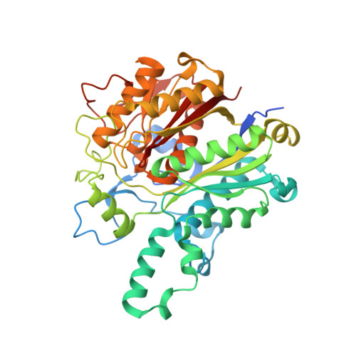
Surface-entropy reduction approaches to manipulate crystal forms of beta-ketoacyl acyl carrier protein synthase II from Streptococcus pneumoniae.
Parthasarathy, G., Cummings, R., Becker, J.W., Soisson, S.M.(2008) Acta Crystallogr D Biol Crystallogr 64: 141-148
- PubMed: 18219113
- DOI: https://doi.org/10.1107/S090744490705559X
- Primary Citation of Related Structures:
2RJT - PubMed Abstract:
A series of experiments with beta-ketoacyl acyl carrier protein synthase II (FabF) from Streptococcus pneumonia (spFabF) were undertaken to evaluate the capability of surface-entropy reduction (SER) to manipulate protein crystallization. Previous work has shown that this protein crystallizes in two forms. The triclinic form contains four molecules in the asymmetric unit (a.u.) and diffracts to 2.1 A resolution, while the more desirable primitive orthorhombic form contains one molecule in the a.u. and diffracts to 1.3 A. The aim was to evaluate the effect of SER mutations that were specifically engineered to avoid perturbing the crystal-packing interfaces employed by the favorable primitive orthorhombic crystal form while potentially disrupting a surface of the protein employed by the less desirable triclinic crystal form. Two mutant proteins were engineered, each of which harbored five SER mutations. Extensive crystallization screening produced crystals of the two mutants, but only under conditions that differed from those used for the native protein. One of the mutant proteins yielded crystals that were of a new form (centered orthorhombic), despite the fact that the interfaces employed by the primitive orthorhombic form of the native protein were specifically unaltered. Structure determination at 1.75 A resolution reveals that one of the mutations, E383A, appears to play a key role in disfavouring the less desirable triclinic crystal form and in generating a new surface for a packing interaction that stabilizes the new crystal form.
- Merck Research Laboratories, Rahway, NJ, USA.
Organizational Affiliation:


