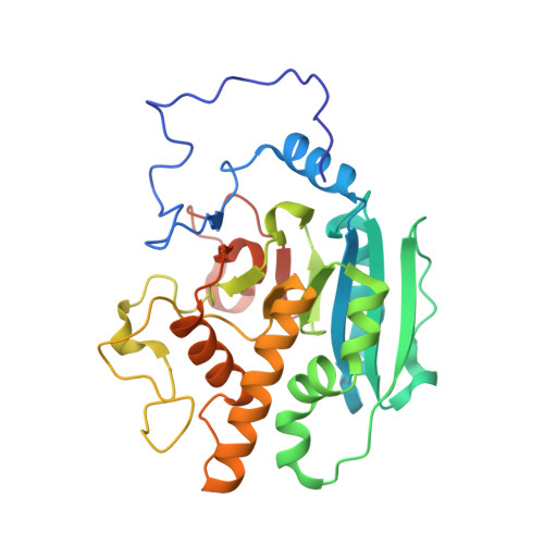
ABO(H) blood group A and B glycosyltransferases recognize substrate via specific conformational changes.
Alfaro, J.A., Zheng, R.B., Persson, M., Letts, J.A., Polakowski, R., Bai, Y., Borisova, S.N., Seto, N.O., Lowary, T.L., Palcic, M.M., Evans, S.V.(2008) J Biological Chem 283: 10097-10108
- PubMed: 18192272
- DOI: https://doi.org/10.1074/jbc.M708669200
- Primary Citation of Related Structures:
2RIT, 2RIX, 2RIY, 2RIZ, 2RJ0, 2RJ1, 2RJ4, 2RJ5, 2RJ6, 2RJ7, 2RJ8, 2RJ9 - PubMed Abstract:
The final step in the enzymatic synthesis of the ABO(H) blood group A and B antigens is catalyzed by two closely related glycosyltransferases, an alpha-(1-->3)-N-acetylgalactosaminyltransferase (GTA) and an alpha-(1-->3)-galactosyltransferase (GTB). Of their 354 amino acid residues, GTA and GTB differ by only four "critical" residues. High resolution structures for GTB and the GTA/GTB chimeric enzymes GTB/G176R and GTB/G176R/G235S bound to a panel of donor and acceptor analog substrates reveal "open," "semi-closed," and "closed" conformations as the enzymes go from the unliganded to the liganded states. In the open form the internal polypeptide loop (amino acid residues 177-195) adjacent to the active site in the unliganded or H antigen-bound enzymes is composed of two alpha-helices spanning Arg(180)-Met(186) and Arg(188)-Asp(194), respectively. The semi-closed and closed forms of the enzymes are generated by binding of UDP or of UDP and H antigen analogs, respectively, and show that these helices merge to form a single distorted helical structure with alternating alpha-3(10)-alpha character that partially occludes the active site. The closed form is distinguished from the semi-closed form by the ordering of the final nine C-terminal residues through the formation of hydrogen bonds to both UDP and H antigen analogs. The semi-closed forms for various mutants generally show significantly more disorder than the open forms, whereas the closed forms display little or no disorder depending strongly on the identity of residue 176. Finally, the use of synthetic analogs reveals how H antigen acceptor binding can be critical in stabilizing the closed conformation. These structures demonstrate a delicately balanced substrate recognition mechanism and give insight on critical aspects of donor and acceptor specificity, on the order of substrate binding, and on the requirements for catalysis.
- Department of Biochemistry and Microbiology, University of Victoria, Victoria, British Columbia, Canada.
Organizational Affiliation:


