2RIP | pdb_00002rip

Structure of DPPIV in complex with an inhibitor


Experimental Data Snapshot

  • Method: X-RAY DIFFRACTION
  • Resolution: 2.90 Å
  • R-Value Free: 
    0.293 (Depositor) 
  • R-Value Work: 
    0.214 (Depositor), 0.208 (DCC) 
  • R-Value Observed: 
    0.218 (Depositor) 

Starting Model: experimental
View more details

wwPDB Validation 3D Report Full Report

Validation slider image for 2RIP

Ligand Structure Quality Assessment 


This is version 2.2 of the entry. See complete history

Literature

Design and synthesis of potent amido- and benzyl-substituted cis-3-amino-4-(2-cyanopyrrolidide)pyrrolidinyl DPP-IV inhibitors.

Corbett, J.W.Dirico, K.Song, W.Boscoe, B.P.Doran, S.D.Boyer, D.Qiu, X.Ammirati, M.Vanvolkenburg, M.A.McPherson, R.K.Parker, J.C.Cox, E.D.

(2007) Bioorg Med Chem Lett 17: 6707-6713

  • DOI: https://doi.org/10.1016/j.bmcl.2007.10.063
  • Primary Citation Related Structures: 
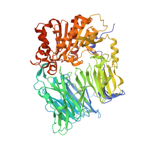
    2RIP

  • PubMed Abstract: 

    The cis-3-amino-4-(2-cyanopyrrolidide)-pyrrolidine template has been shown to afford low nanomolar inhibitors of human DPP-IV that exhibit a robust PK/PD profile. An X-ray co-crystal structure of 5 confirmed the proposed mode of binding. The potent single digit DPP-IV inhibitor 53 exhibited a preferred PK/PD profile in preclinical animal models and was selected for additional profiling.


  • Organizational Affiliation
    • Pfizer Global Research and Development, Eastern Point Road, Groton, CT 06340, USA.

Macromolecule Content 

  • Total Structure Weight: 86.98 kDa 
  • Atom Count: 6,158 
  • Modeled Residue Count: 729 
  • Deposited Residue Count: 729 
  • Unique protein chains: 1

Macromolecules

Find similar proteins by:|  3D Structure
Entity ID: 1
MoleculeChains  Sequence LengthOrganismDetailsImage
Dipeptidyl peptidase 4729Homo sapiensMutation(s): 0 
Gene Names: DPPIV
EC: 3.4.14.5
UniProt & NIH Common Fund Data Resources
Find proteins for P27487 (Homo sapiens)
Explore P27487 
Go to UniProtKB:  P27487
PHAROS:  P27487
GTEx:  ENSG00000197635 
Entity Groups
Sequence Clusters30% Identity50% Identity70% Identity90% Identity95% Identity100% Identity
UniProt GroupP27487
Glycosylation
Glycosylation Sites: 6Go to GlyGen: P27487-1
Sequence Annotations
Expand
Reference Sequence

Oligosaccharides

Help  
Entity ID: 2
MoleculeChains Length2D Diagram GlycosylationD Interactions
alpha-D-mannopyranose-(1-6)-alpha-D-mannopyranose-(1-4)-2-acetamido-2-deoxy-beta-D-glucopyranose-(1-4)-2-acetamido-2-deoxy-beta-D-glucopyranose
B
4N-Glycosylation
Glycosylation Resources
GlyTouCan: G04854NQ
GlyCosmos: G04854NQ
GlyGen: G04854NQ
Entity ID: 3
MoleculeChains Length2D Diagram GlycosylationD Interactions
2-acetamido-2-deoxy-beta-D-glucopyranose-(1-4)-2-acetamido-2-deoxy-beta-D-glucopyranose
C
2N-Glycosylation
Glycosylation Resources
GlyTouCan: G42666HT
GlyCosmos: G42666HT
GlyGen: G42666HT

Experimental Data & Validation

Experimental Data

  • Method: X-RAY DIFFRACTION
  • Resolution: 2.90 Å
  • R-Value Free:  0.293 (Depositor) 
  • R-Value Work:  0.214 (Depositor), 0.208 (DCC) 
  • R-Value Observed: 0.218 (Depositor) 
Space Group: P 43 21 2
Unit Cell:
Length ( Å )Angle ( ˚ )
a = 69.22α = 90
b = 69.22β = 90
c = 409.22γ = 90
Software Package:
Software NamePurpose
REFMACrefinement
HKL-2000data collection
HKL-2000data reduction
HKL-2000data scaling
AMoREphasing

Structure Validation

View Full Validation Report


Ligand Structure Quality Assessment 


Entry History 

Deposition Data

  • Released Date: 2008-03-18 
  • Deposition Author(s): Qiu, X.

Revision History  (Full details and data files)

  • Version 1.0: 2008-03-18
    Type: Initial release
  • Version 1.1: 2011-07-13
    Changes: Non-polymer description, Version format compliance
  • Version 2.0: 2020-07-29
    Type: Remediation
    Reason: Carbohydrate remediation
    Changes: Advisory, Atomic model, Data collection, Derived calculations, Structure summary
  • Version 2.1: 2023-08-30
    Changes: Data collection, Database references, Refinement description, Structure summary
  • Version 2.2: 2024-11-20
    Changes: Structure summary