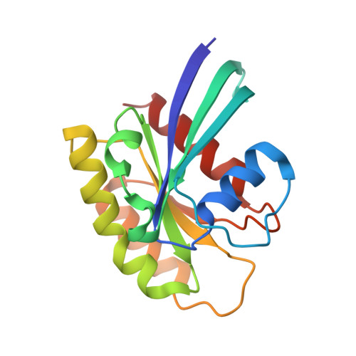
RCSB PDB - 2RGB: Crystal structure of H-RasQ61K-GppNHp

 2RGB

Crystal structure of H-RasQ61K-GppNHp


Experimental Data Snapshot

  • Method: X-RAY DIFFRACTION
  • Resolution: 1.35 Å
  • R-Value Free: 0.218 
  • R-Value Work: 0.200 
  • R-Value Observed: 0.200 

Starting Model: experimental
View more details

wwPDB Validation   3D Report Full Report


Ligand Structure Quality Assessment 

Created with Raphaël 2.3.0Worse 01 BetterLigand structure goodness of fit to experimental dataBest fitted GNPClick on this verticalbar to view details

This is version 1.3 of the entry. See complete history


Literature

Transformation Efficiency of RasQ61 Mutants Linked to Structural Features of the Switch Regions in the Presence of Raf.

Buhrman, G.Wink, G.Mattos, C.

(2007) Structure 15: 1618-1629

  • DOI: https://doi.org/10.1016/j.str.2007.10.011
  • Primary Citation of Related Structures:  
    2RGA, 2RGB, 2RGC, 2RGD, 2RGE, 2RGG

  • PubMed Abstract: 

    Transformation efficiencies of Ras mutants at residue 61 range over three orders of magnitude, but the in vitro GTPase activity decreases 10-fold for all mutants. We show that Raf impairs the GTPase activity of RasQ61L, suggesting that the Ras/Raf complex differentially modulates transformation. Our crystal structures show that, in transforming mutants, switch II takes part in a network of hydrophobic interactions burying the nucleotide and precatalytic water molecule. Our results suggest that Y32 and a water molecule bridging it to the gamma-phosphate in the wild-type structure play a role in GTP hydrolysis in lieu of the Arg finger in the absence of GAP. The bridging water molecule is absent in the transforming mutants, contributing to the burying of the nucleotide. We propose a mechanism for intrinsic hydrolysis in Raf-bound Ras and elucidate structural features in the Q61 mutants that correlate with their potency to transform cells.


  • Organizational Affiliation

    Department of Molecular and Structural Biochemistry, North Carolina State University, 128 Polk Hall-CB 7622, Raleigh, NC 27695, USA.


Macromolecules
Find similar proteins by:  (by identity cutoff)  |  3D Structure
Entity ID: 1
MoleculeChains Sequence LengthOrganismDetailsImage
GTPase HRas166Homo sapiensMutation(s): 1 
Gene Names: HRASHRAS1
EC: 3.6.5.2
UniProt & NIH Common Fund Data Resources
Find proteins for P01112 (Homo sapiens)
Explore P01112 
Go to UniProtKB:  P01112
PHAROS:  P01112
GTEx:  ENSG00000174775 
Entity Groups  
Sequence Clusters30% Identity50% Identity70% Identity90% Identity95% Identity100% Identity
UniProt GroupP01112
Sequence Annotations
Expand
  • Reference Sequence
Experimental Data & Validation

Experimental Data

  • Method: X-RAY DIFFRACTION
  • Resolution: 1.35 Å
  • R-Value Free: 0.218 
  • R-Value Work: 0.200 
  • R-Value Observed: 0.200 
  • Space Group: H 3 2
Unit Cell:
Length ( Å )Angle ( ˚ )
a = 88.695α = 90
b = 88.695β = 90
c = 133.529γ = 120
Software Package:
Software NamePurpose
CNSrefinement
HKL-2000data collection
HKL-2000data reduction
HKL-2000data scaling
CNSphasing

Structure Validation

View Full Validation Report


Ligand Structure Quality Assessment 

Created with Raphaël 2.3.0Worse 01 BetterLigand structure goodness of fit to experimental dataBest fitted GNPClick on this verticalbar to view details

Entry History 

Deposition Data

Revision History  (Full details and data files)

  • Version 1.0: 2007-12-18
    Type: Initial release
  • Version 1.1: 2011-07-13
    Changes: Version format compliance
  • Version 1.2: 2021-10-20
    Changes: Database references, Derived calculations
  • Version 1.3: 2023-08-30
    Changes: Data collection, Refinement description