


Crystal structure of the effector domain of PLXNB1 bound with Rnd1 GTPase.
Tong, Y., Tempel, W., Shen, L., Arrowsmith, C.H., Edwards, A.M., Sundstrom, M., Weigelt, J., Bochkarev, A., Park, H.To be published.
Experimental Data Snapshot
Starting Models: experimental
View more details
Entity ID: 1 | |||||
|---|---|---|---|---|---|
| Molecule | Chains | Sequence Length | Organism | Details | Image |
| Plexin-B1 | 121 | Homo sapiens | Mutation(s): 0 Gene Names: PLXNB1, KIAA0407, PLXN5, SEP | 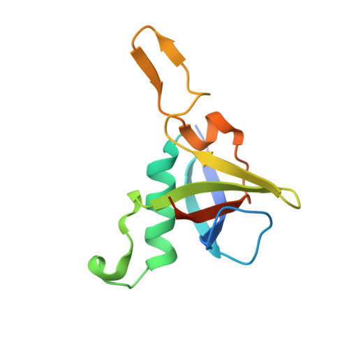 | |
UniProt & NIH Common Fund Data Resources | |||||
PHAROS: O43157 GTEx: ENSG00000164050 | |||||
Entity Groups | |||||
| Sequence Clusters | 30% Identity50% Identity70% Identity90% Identity95% Identity100% Identity | ||||
| UniProt Group | O43157 | ||||
Sequence AnnotationsExpand | |||||
Reference Sequence | |||||
Entity ID: 2 | |||||
|---|---|---|---|---|---|
| Molecule | Chains | Sequence Length | Organism | Details | Image |
| Rho-related GTP-binding protein Rho6 | 197 | Homo sapiens | Mutation(s): 0 Gene Names: RND1, RHO6 EC: 3.6.5.2 | 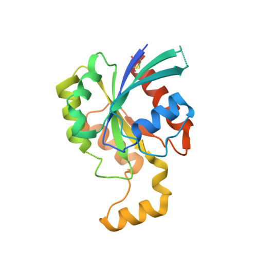 | |
UniProt & NIH Common Fund Data Resources | |||||
PHAROS: Q92730 GTEx: ENSG00000172602 | |||||
Entity Groups | |||||
| Sequence Clusters | 30% Identity50% Identity70% Identity90% Identity95% Identity100% Identity | ||||
| UniProt Group | Q92730 | ||||
Sequence AnnotationsExpand | |||||
Reference Sequence | |||||
| Ligands 4 Unique | |||||
|---|---|---|---|---|---|
| ID | Chains | Name / Formula / InChI Key | 2D Diagram | 3D Interactions | |
| GNP Download:Ideal Coordinates CCD File | DA [auth D], K [auth B] | PHOSPHOAMINOPHOSPHONIC ACID-GUANYLATE ESTER C10 H17 N6 O13 P3 UQABYHGXWYXDTK-UUOKFMHZSA-N |  | ||
| CA Download:Ideal Coordinates CCD File | CA [auth D], J [auth B] | CALCIUM ION Ca BHPQYMZQTOCNFJ-UHFFFAOYSA-N |  | ||
| MG Download:Ideal Coordinates CCD File | BA [auth D], I [auth B] | MAGNESIUM ION Mg JLVVSXFLKOJNIY-UHFFFAOYSA-N |  | ||
| UNX Download:Ideal Coordinates CCD File | AA [auth C] E [auth A] EA [auth D] F [auth A] FA [auth D] | UNKNOWN ATOM OR ION X |  | ||
| Length ( Å ) | Angle ( ˚ ) |
|---|---|
| a = 150.136 | α = 90 |
| b = 71.653 | β = 128.36 |
| c = 101.885 | γ = 90 |
| Software Name | Purpose |
|---|---|
| DENZO | data reduction |
| SCALEPACK | data scaling |
| PHASER | phasing |
| REFMAC | refinement |
| PDB_EXTRACT | data extraction |
| ADSC | data collection |
| HKL-2000 | data reduction |
| HKL-2000 | data scaling |


