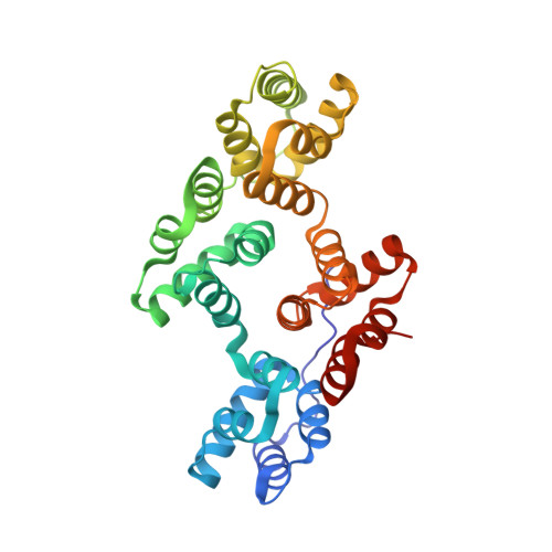
2RAN

RAT ANNEXIN V CRYSTAL STRUCTURE: CA2+-INDUCED CONFORMATIONAL CHANGES


Experimental Data Snapshot

  • Method: X-RAY DIFFRACTION
  • Resolution: 1.89 Å
  • R-Value Work: 0.212 
  • R-Value Observed: 0.212 

wwPDB Validation   3D Report Full Report


This is version 1.3 of the entry. See complete history


Literature

Rat annexin V crystal structure: Ca(2+)-induced conformational changes.

Concha, N.O.Head, J.F.Kaetzel, M.A.Dedman, J.R.Seaton, B.A.

(1993) Science 261: 1321-1324

  • DOI: https://doi.org/10.1126/science.8362244
  • Primary Citation of Related Structures:  
    2RAN

  • PubMed Abstract: 

    Annexins are a family of calcium- and phospholipid-binding proteins implicated in mediating membrane-related processes such as secretion, signal transduction, and ion channel activity. The crystal structure of rat annexin V was solved to 1.9 angstrom resolution by multiple isomorphous replacement. Unlike previously solved annexin V structures, all four domains bound calcium in this structure. Calcium binding in the third domain induced a large relocation of the calcium-binding loop regions, exposing the single tryptophan residue to the solvent. These alterations in annexin V suggest a role for domain 3 in calcium-triggered interaction with phospholipid membranes.


  • Organizational Affiliation

    Department of Physiology, Boston University School of Medicine, MA 02118.


Macromolecules
Find similar proteins by:  (by identity cutoff)  |  3D Structure
Entity ID: 1
MoleculeChains Sequence LengthOrganismDetailsImage
ANNEXIN V316Rattus norvegicusMutation(s): 0 
UniProt
Find proteins for P14668 (Rattus norvegicus)
Explore P14668 
Go to UniProtKB:  P14668
Entity Groups  
Sequence Clusters30% Identity50% Identity70% Identity90% Identity95% Identity100% Identity
UniProt GroupP14668
Sequence Annotations
Expand
  • Reference Sequence
Small Molecules
Experimental Data & Validation

Experimental Data

  • Method: X-RAY DIFFRACTION
  • Resolution: 1.89 Å
  • R-Value Work: 0.212 
  • R-Value Observed: 0.212 
  • Space Group: H 3
Unit Cell:
Length ( Å )Angle ( ˚ )
a = 156.9α = 90
b = 156.9β = 90
c = 37.03γ = 120
Software Package:
Software NamePurpose
X-PLORmodel building
X-PLORrefinement
X-PLORphasing

Structure Validation

View Full Validation Report


Entry History 

Deposition Data

Revision History  (Full details and data files)

  • Version 1.0: 1994-11-30
    Type: Initial release
  • Version 1.1: 2008-03-03
    Changes: Version format compliance
  • Version 1.2: 2011-07-13
    Changes: Version format compliance
  • Version 1.3: 2024-02-21
    Changes: Data collection, Database references, Derived calculations, Other