2R9K | pdb_00002r9k

Crystal Structure of Misteltoe Lectin I in Complex with Phloretamide


Experimental Data Snapshot

  • Method: X-RAY DIFFRACTION
  • Resolution: 2.70 Å
  • R-Value Free: 
    0.267 (Depositor), 0.236 (DCC) 
  • R-Value Work: 
    0.226 (Depositor), 0.225 (DCC) 
  • R-Value Observed: 
    0.228 (Depositor) 

Starting Model: experimental
View more details

wwPDB Validation 3D Report Full Report

Validation slider image for 2R9K

Ligand Structure Quality Assessment 


This is version 3.2 of the entry. See complete history

Literature

The mistletoe lectin I--phloretamide structure reveals a new function of plant lectins.

Meyer, A.Rypniewski, W.Celewicz, L.Erdmann, V.A.Voelter, W.Singh, T.P.Genov, N.Barciszewski, J.Betzel, C.h.

(2007) Biochem Biophys Res Commun 364: 195-200

  • DOI: https://doi.org/10.1016/j.bbrc.2007.09.113
  • Primary Citation Related Structures: 
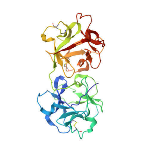
    2R9K

  • PubMed Abstract: 

    The X-ray structure at 2.7A resolution of the complex between the European mistletoe lectin I (Viscum album, ML-I) and the plant growth hormone, 3-(p-hydroxyphenyl)-propionic acid amide (phloretamide, PA) from xylem sap has revealed the binding of PA at the so far undescribed hydrophobic cavity located between the two subunits of this ribosome-inhibiting protein. No such cavity is observed in related lectins. The binding of PA is achieved through interactions with the non-conserved residues Val228A, Leu230A, Arg388B, and the C-terminal Pro510B. It is conceivable that binding of PA to ML-I is part of a defence mechanism of the parasite against the host, whereby the parasite prevents the growth hormone of the host from interfering with its own regulatory system. The specific binding of PA to ML-I indicates that heterodimeric RIPs are multifunctional proteins whose functions in the cell have not yet been fully recognized and analyzed.


  • Organizational Affiliation
    • Institute of Biochemistry and Molecular Biology, University of Hamburg, c/o DESY, Notkestr. 85, Building 22a, 22603 Hamburg, Germany.

Macromolecule Content 

  • Total Structure Weight: 58.65 kDa 
  • Atom Count: 4,135 
  • Modeled Residue Count: 510 
  • Deposited Residue Count: 517 
  • Unique protein chains: 2

Macromolecules

Find similar proteins by:|  3D Structure
Entity ID: 1
MoleculeChains  Sequence LengthOrganismDetailsImage
Beta-galactoside-specific lectin 1254Viscum albumMutation(s): 0 
UniProt
Find proteins for P81446 (Viscum album)
Explore P81446 
Go to UniProtKB:  P81446
Entity Groups
Sequence Clusters30% Identity50% Identity70% Identity90% Identity95% Identity100% Identity
UniProt GroupP81446
Sequence Annotations
Expand
Reference Sequence
Find similar proteins by:|  3D Structure
Entity ID: 2
MoleculeChains  Sequence LengthOrganismDetailsImage
Beta-galactoside-specific lectin 1 chain B263Viscum albumMutation(s): 0 
UniProt
Find proteins for P81446 (Viscum album)
Explore P81446 
Go to UniProtKB:  P81446
Entity Groups
Sequence Clusters30% Identity50% Identity70% Identity90% Identity95% Identity100% Identity
UniProt GroupP81446
Glycosylation
Glycosylation Sites: 2
Sequence Annotations
Expand
Reference Sequence

Oligosaccharides

Help  
Entity ID: 3
MoleculeChains Length2D Diagram GlycosylationD Interactions
2-acetamido-2-deoxy-beta-D-glucopyranose-(4-4)-2-acetamido-2-deoxy-beta-D-glucopyranose
C
2N/AN-Glycosylation

Small Molecules

Ligands 5 Unique
IDChains Name / Formula / InChI Key2D Diagram3D Interactions
NAG

Query on NAG


Download:Ideal Coordinates CCD File
D [auth A],
L [auth B],
M [auth B],
N [auth B]
2-acetamido-2-deoxy-beta-D-glucopyranose
C8 H15 N O6
OVRNDRQMDRJTHS-FMDGEEDCSA-N
SGI

Query on SGI


Download:Ideal Coordinates CCD File
K [auth A]3-(4-hydroxyphenyl)propanamide
C9 H11 N O2
OEHZEBOCZWCVMK-UHFFFAOYSA-N
SO4

Query on SO4


Download:Ideal Coordinates CCD File
E [auth A],
F [auth A],
G [auth A],
O [auth B]
SULFATE ION
O4 S
QAOWNCQODCNURD-UHFFFAOYSA-L
GOL

Query on GOL


Download:Ideal Coordinates CCD File
H [auth A],
I [auth A],
P [auth B],
Q [auth B]
GLYCEROL
C3 H8 O3
PEDCQBHIVMGVHV-UHFFFAOYSA-N
CL

Query on CL


Download:Ideal Coordinates CCD File
J [auth A]CHLORIDE ION
Cl
VEXZGXHMUGYJMC-UHFFFAOYSA-M

Experimental Data & Validation

Experimental Data

  • Method: X-RAY DIFFRACTION
  • Resolution: 2.70 Å
  • R-Value Free:  0.267 (Depositor), 0.236 (DCC) 
  • R-Value Work:  0.226 (Depositor), 0.225 (DCC) 
  • R-Value Observed: 0.228 (Depositor) 
Space Group: P 65 2 2
Unit Cell:
Length ( Å )Angle ( ˚ )
a = 107.059α = 90
b = 107.059β = 90
c = 312.375γ = 120
Software Package:
Software NamePurpose
REFMACrefinement
MAR345dtbdata collection
DENZOdata reduction
SCALEPACKdata scaling
MOLREPphasing

Structure Validation

View Full Validation Report


Ligand Structure Quality Assessment 


Entry History 

Deposition Data

Revision History  (Full details and data files)

  • Version 1.0: 2007-10-30
    Type: Initial release
  • Version 1.1: 2011-07-13
    Changes: Non-polymer description, Version format compliance
  • Version 2.0: 2020-07-29
    Type: Remediation
    Reason: Carbohydrate remediation
    Changes: Advisory, Atomic model, Data collection, Derived calculations, Structure summary
  • Version 2.1: 2022-04-20
    Changes: Database references, Derived calculations, Source and taxonomy, Structure summary
  • Version 3.0: 2022-05-04
    Changes: Advisory, Atomic model, Data collection, Derived calculations, Structure summary
  • Version 3.1: 2023-08-30
    Changes: Data collection, Refinement description
  • Version 3.2: 2024-11-20
    Changes: Structure summary