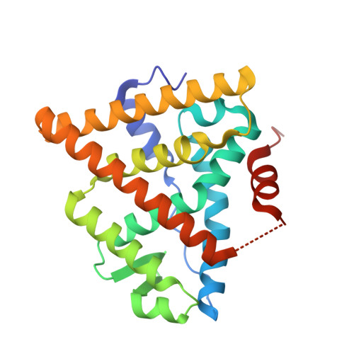
2R6W

Estrogen receptor alpha ligand-binding domain complexed to a SERM


Experimental Data Snapshot

  • Method: X-RAY DIFFRACTION
  • Resolution: 2.00 Å
  • R-Value Free: 0.257 
  • R-Value Work: 0.208 
  • R-Value Observed: 0.212 

Starting Model: experimental
View more details

wwPDB Validation   3D Report Full Report


Ligand Structure Quality Assessment 


This is version 1.3 of the entry. See complete history


Literature

Prediction of the tissue-specificity of selective estrogen receptor modulators by using a single biochemical method.

Dai, S.Y.Chalmers, M.J.Bruning, J.Bramlett, K.S.Osborne, H.E.Montrose-Rafizadeh, C.Barr, R.J.Wang, Y.Wang, M.Burris, T.P.Dodge, J.A.Griffin, P.R.

(2008) Proc Natl Acad Sci U S A 105: 7171-7176

  • DOI: https://doi.org/10.1073/pnas.0710802105
  • Primary Citation of Related Structures:  
    2R6W, 2R6Y

  • PubMed Abstract: 

    Here, we demonstrate that a single biochemical assay is able to predict the tissue-selective pharmacology of an array of selective estrogen receptor modulators (SERMs). We describe an approach to classify estrogen receptor (ER) modulators based on dynamics of the receptor-ligand complex as probed with hydrogen/deuterium exchange (HDX) mass spectrometry. Differential HDX mapping coupled with cluster and discriminate analysis effectively predicted tissue-selective function in most, but not all, cases tested. We demonstrate that analysis of dynamics of the receptor-ligand complex facilitates binning of ER modulators into distinct groups based on structural dynamics. Importantly, we were able to differentiate small structural changes within ER ligands of the same chemotype. In addition, HDX revealed differentially stabilized regions within the ligand-binding pocket that may contribute to the different pharmacology phenotypes of the compounds independent of helix 12 positioning. In summary, HDX provides a sensitive and rapid approach to classify modulators of the estrogen receptor that correlates with their pharmacological profile.


  • Organizational Affiliation

    Department of Molecular Therapeutics, The Scripps Research Institute, Jupiter, FL 33458, USA.


Macromolecules
Find similar proteins by:  (by identity cutoff)  |  3D Structure
Entity ID: 1
MoleculeChains Sequence LengthOrganismDetailsImage
Estrogen receptor
A, B
248Homo sapiensMutation(s): 3 
Gene Names: ESR1ESRNR3A1
UniProt & NIH Common Fund Data Resources
Find proteins for P03372 (Homo sapiens)
Explore P03372 
Go to UniProtKB:  P03372
PHAROS:  P03372
GTEx:  ENSG00000091831 
Entity Groups  
Sequence Clusters30% Identity50% Identity70% Identity90% Identity95% Identity100% Identity
UniProt GroupP03372
Sequence Annotations
Expand
  • Reference Sequence
Small Molecules
Ligands 1 Unique
IDChains Name / Formula / InChI Key2D Diagram3D Interactions
LLB
Query on LLB

Download Ideal Coordinates CCD File 
C [auth A],
D [auth B]
[6-hydroxy-2-(4-hydroxyphenyl)-1-benzothien-3-yl]{4-[2-(4-methylpiperidin-1-yl)ethoxy]phenyl}methanone
C29 H29 N O4 S
RLOZWABDAGQFIY-UHFFFAOYSA-N
Binding Affinity Annotations 
IDSourceBinding Affinity
LLB PDBBind:  2R6W Ki: 0.44 (nM) from 1 assay(s)
Experimental Data & Validation

Experimental Data

  • Method: X-RAY DIFFRACTION
  • Resolution: 2.00 Å
  • R-Value Free: 0.257 
  • R-Value Work: 0.208 
  • R-Value Observed: 0.212 
  • Space Group: C 2 2 21
Unit Cell:
Length ( Å )Angle ( ˚ )
a = 56.47α = 90
b = 102.761β = 90
c = 172.952γ = 90
Software Package:
Software NamePurpose
REFMACrefinement
MAR345dtbdata collection
HKL-2000data reduction
HKL-2000data scaling
AMoREphasing

Structure Validation

View Full Validation Report


Ligand Structure Quality Assessment 


Entry History 

Deposition Data

  • Released Date: 2008-04-08 
  • Deposition Author(s): Wang, Y.

Revision History  (Full details and data files)

  • Version 1.0: 2008-04-08
    Type: Initial release
  • Version 1.1: 2011-07-13
    Changes: Version format compliance
  • Version 1.2: 2021-10-20
    Changes: Database references, Derived calculations
  • Version 1.3: 2023-08-30
    Changes: Data collection, Refinement description