2R5E

Aedes kynurenine aminotransferase in complex with glutamine


Experimental Data Snapshot

  • Method: X-RAY DIFFRACTION
  • Resolution: 1.84 Å
  • R-Value Free: 0.243 
  • R-Value Work: 0.201 
  • R-Value Observed: 0.203 

wwPDB Validation   3D Report Full Report


Ligand Structure Quality Assessment 


This is version 1.2 of the entry. See complete history


Literature

Structural insight into the mechanism of substrate specificity of aedes kynurenine aminotransferase.

Han, Q.Gao, Y.G.Robinson, H.Li, J.

(2008) Biochemistry 47: 1622-1630

  • DOI: https://doi.org/10.1021/bi701800j
  • Primary Citation of Related Structures:  
    2R5C, 2R5E

  • PubMed Abstract: 

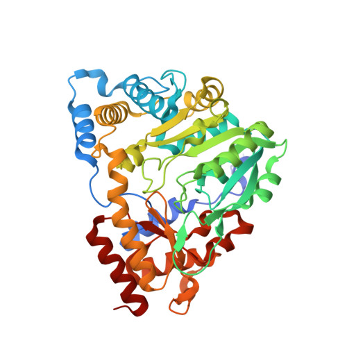
    Aedes aegypti kynurenine aminotransferase (AeKAT) is a multifunctional aminotransferase. It catalyzes the transamination of a number of amino acids and uses many biologically relevant alpha-keto acids as amino group acceptors. AeKAT also is a cysteine S-conjugate beta-lyase. The most important function of AeKAT is the biosynthesis of kynurenic acid, a natural antagonist of NMDA and alpha7-nicotinic acetylcholine receptors. Here, we report the crystal structures of AeKAT in complex with its best amino acid substrates, glutamine and cysteine. Glutamine is found in both subunits of the biological dimer, and cysteine is found in one of the two subunits. Both substrates form external aldemines with pyridoxal 5-phosphate in the structures. This is the first instance in which one pyridoxal 5-phosphate enzyme has been crystallized with cysteine or glutamine forming external aldimine complexes, cysteinyl aldimine and glutaminyl aldimine. All the units with substrate are in the closed conformation form, and the unit without substrate is in the open form, which suggests that the binding of substrate induces the conformation change of AeKAT. By comparing the active site residues of the AeKAT-cysteine structure with those of the human KAT I-phenylalanine structure, we determined that Tyr286 in AeKAT is changed to Phe278 in human KAT I, which may explain why AeKAT transaminates hydrophilic amino acids more efficiently than human KAT I does.


  • Organizational Affiliation

    Department of Biochemistry, Virginia Tech, Blacksburg, Virginia 24061, USA.


Macromolecules
Find similar proteins by:  (by identity cutoff)  |  3D Structure
Entity ID: 1
MoleculeChains Sequence LengthOrganismDetailsImage
Kynurenine aminotransferase
A, B
429Aedes aegyptiMutation(s): 0 
Gene Names: KAT
EC: 2.6.1.7 (PDB Primary Data), 2.6.1.64 (PDB Primary Data)
UniProt
Find proteins for Q17CS8 (Aedes aegypti)
Explore Q17CS8 
Go to UniProtKB:  Q17CS8
Entity Groups  
Sequence Clusters30% Identity50% Identity70% Identity90% Identity95% Identity100% Identity
UniProt GroupQ17CS8
Sequence Annotations
Expand
  • Reference Sequence
Small Molecules
Ligands 1 Unique
IDChains Name / Formula / InChI Key2D Diagram3D Interactions
QLP
Query on QLP

Download Ideal Coordinates CCD File 
C [auth A],
D [auth B]
N~2~-({3-HYDROXY-2-METHYL-5-[(PHOSPHONOOXY)METHYL]PYRIDIN-4-YL}METHYL)-L-GLUTAMINE
C13 H20 N3 O8 P
TUUVENFNODJVKV-JTQLQIEISA-N
Experimental Data & Validation

Experimental Data

  • Method: X-RAY DIFFRACTION
  • Resolution: 1.84 Å
  • R-Value Free: 0.243 
  • R-Value Work: 0.201 
  • R-Value Observed: 0.203 
  • Space Group: P 21 21 21
Unit Cell:
Length ( Å )Angle ( ˚ )
a = 56.374α = 90
b = 95.589β = 90
c = 165.901γ = 90
Software Package:
Software NamePurpose
REFMACrefinement
HKL-2000data collection
HKL-2000data reduction
HKL-2000data scaling
MOLREPphasing

Structure Validation

View Full Validation Report


Ligand Structure Quality Assessment 


Entry History 

Deposition Data

Revision History  (Full details and data files)

  • Version 1.0: 2008-03-18
    Type: Initial release
  • Version 1.1: 2011-07-13
    Changes: Version format compliance
  • Version 1.2: 2023-08-30
    Changes: Data collection, Database references, Derived calculations, Refinement description