2R5B | pdb_00002r5b

Structure of the gp41 N-trimer in complex with the HIV entry inhibitor PIE7


Experimental Data Snapshot

  • Method: X-RAY DIFFRACTION
  • Resolution: 2.00 Å
  • R-Value Free: 
    0.279 (Depositor), 0.278 (DCC) 
  • R-Value Work: 
    0.203 (Depositor), 0.217 (DCC) 
  • R-Value Observed: 
    0.209 (Depositor) 

wwPDB Validation 3D Report Full Report

Validation slider image for 2R5B

This is version 1.4 of the entry. See complete history

Literature

Potent D-peptide inhibitors of HIV-1 entry

Welch, B.D.Vandemark, A.P.Heroux, A.Hill, C.P.Kay, M.S.

(2007) Proc Natl Acad Sci U S A 104: 16828-16833

  • DOI: https://doi.org/10.1073/pnas.0708109104
  • Primary Citation Related Structures: 
    2R3C, 2R5B, 2R5D

  • PubMed Abstract: 

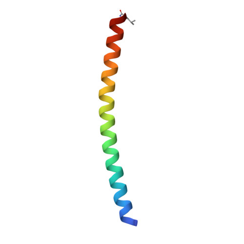
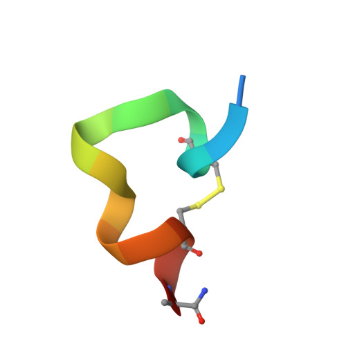
    During HIV-1 entry, the highly conserved gp41 N-trimer pocket region becomes transiently exposed and vulnerable to inhibition. Using mirror-image phage display and structure-assisted design, we have discovered protease-resistant D-amino acid peptides (D-peptides) that bind the N-trimer pocket with high affinity and potently inhibit viral entry. We also report high-resolution crystal structures of two of these D-peptides in complex with a pocket mimic that suggest sources of their high potency. A trimeric version of one of these peptides is the most potent pocket-specific entry inhibitor yet reported by three orders of magnitude (IC(50) = 250 pM). These results are the first demonstration that D-peptides can form specific and high-affinity interactions with natural protein targets and strengthen their promise as therapeutic agents. The D-peptides described here address limitations associated with current L-peptide entry inhibitors and are promising leads for the prevention and treatment of HIV/AIDS.


  • Organizational Affiliation
    • Department of Biochemistry, University of Utah, Emma Eccles Jones Medical Research Building, 15 North Medical Drive East, Salt Lake City, UT 84112-5650, USA.

Macromolecule Content 

  • Total Structure Weight: 21.98 kDa 
  • Atom Count: 1,671 
  • Modeled Residue Count: 191 
  • Deposited Residue Count: 192 
  • Unique protein chains: 2

Macromolecules

Find similar proteins by:|  3D Structure
Entity ID: 1
MoleculeChains  Sequence LengthOrganismDetailsImage
gp41 N-peptide
A, B, C
47synthetic constructMutation(s): 0 
Find similar proteins by:  Sequence   |   3D Structure  
Entity ID: 2
MoleculeChains  Sequence LengthOrganismDetailsImage
HIV entry inhibitor PIE7D [auth H],
E [auth K],
F [auth L]
17synthetic constructMutation(s): 0 

Experimental Data & Validation

Experimental Data

  • Method: X-RAY DIFFRACTION
  • Resolution: 2.00 Å
  • R-Value Free:  0.279 (Depositor), 0.278 (DCC) 
  • R-Value Work:  0.203 (Depositor), 0.217 (DCC) 
  • R-Value Observed: 0.209 (Depositor) 
Space Group: C 2 2 21
Unit Cell:
Length ( Å )Angle ( ˚ )
a = 47.024α = 90
b = 106.201β = 90
c = 76.7γ = 90
Software Package:
Software NamePurpose
DENZOdata reduction
SCALEPACKdata scaling
PHASERphasing
REFMACrefinement
PDB_EXTRACTdata extraction
CrystalCleardata collection

Structure Validation

View Full Validation Report


Entry History 

Deposition Data

Revision History  (Full details and data files)

  • Version 1.0: 2007-10-02
    Type: Initial release
  • Version 1.1: 2011-07-13
    Changes: Atomic model, Database references, Derived calculations, Non-polymer description, Structure summary, Version format compliance
  • Version 1.2: 2012-12-12
    Changes: Other
  • Version 1.3: 2017-10-25
    Changes: Refinement description, Source and taxonomy
  • Version 1.4: 2024-11-13
    Changes: Data collection, Database references, Derived calculations, Structure summary