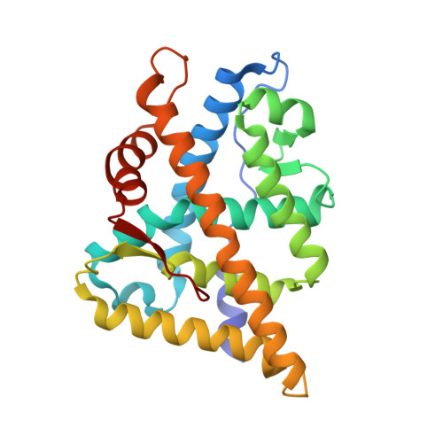
2QPY

AR LBD with small molecule


Experimental Data Snapshot

  • Method: X-RAY DIFFRACTION
  • Resolution: 2.50 Å
  • R-Value Free: 0.269 
  • R-Value Work: 0.269 
  • R-Value Observed: 0.269 

wwPDB Validation   3D Report Full Report


Ligand Structure Quality Assessment 


This is version 1.3 of the entry. See complete history


Literature

A surface on the androgen receptor that allosterically regulates coactivator binding.

Estebanez-Perpina, E.Arnold, L.A.Arnold, A.A.Nguyen, P.Rodrigues, E.D.Mar, E.Bateman, R.Pallai, P.Shokat, K.M.Baxter, J.D.Guy, R.K.Webb, P.Fletterick, R.J.

(2007) Proc Natl Acad Sci U S A 104: 16074-16079

  • DOI: https://doi.org/10.1073/pnas.0708036104
  • Primary Citation of Related Structures:  
    2PIO, 2PIP, 2PIQ, 2PIR, 2PIT, 2PIU, 2PIV, 2PIW, 2PIX, 2PKL, 2QPY

  • PubMed Abstract: 

    Current approaches to inhibit nuclear receptor (NR) activity target the hormone binding pocket but face limitations. We have proposed that inhibitors, which bind to nuclear receptor surfaces that mediate assembly of the receptor's binding partners, might overcome some of these limitations. The androgen receptor (AR) plays a central role in prostate cancer, but conventional inhibitors lose effectiveness as cancer treatments because anti-androgen resistance usually develops. We conducted functional and x-ray screens to identify compounds that bind the AR surface and block binding of coactivators for AR activation function 2 (AF-2). Four compounds that block coactivator binding in solution with IC(50) approximately 50 microM and inhibit AF-2 activity in cells were detected: three nonsteroidal antiinflammatory drugs and the thyroid hormone 3,3',5-triiodothyroacetic acid. Although visualization of compounds at the AR surface reveals weak binding at AF-2, the most potent inhibitors bind preferentially to a previously unknown regulatory surface cleft termed binding function (BF)-3, which is a known target for mutations in prostate cancer and androgen insensitivity syndrome. X-ray structural analysis reveals that 3,3',5-triiodothyroacetic acid binding to BF-3 remodels the adjacent interaction site AF-2 to weaken coactivator binding. Mutation of residues that form BF-3 inhibits AR function and AR AF-2 activity. We propose that BF-3 is a previously unrecognized allosteric regulatory site needed for AR activity in vivo and a possible pharmaceutical target.


  • Organizational Affiliation

    Department of Biochemistry and Biophysics, University of California, San Francisco, CA 94143, USA.


Macromolecules
Find similar proteins by:  (by identity cutoff)  |  3D Structure
Entity ID: 1
MoleculeChains Sequence LengthOrganismDetailsImage
Androgen receptor251Mus musculusMutation(s): 0 
Gene Names: ArNr3c4
UniProt
Find proteins for P19091 (Mus musculus)
Explore P19091 
Go to UniProtKB:  P19091
Entity Groups  
Sequence Clusters30% Identity50% Identity70% Identity90% Identity95% Identity100% Identity
UniProt GroupP19091
Sequence Annotations
Expand
  • Reference Sequence

Find similar proteins by:  Sequence   |   3D Structure  

Entity ID: 2
MoleculeChains Sequence LengthOrganismDetailsImage
Coactivator peptide10synthetic constructMutation(s): 0 
UniProt & NIH Common Fund Data Resources
Find proteins for Q61026 (Mus musculus)
Explore Q61026 
Go to UniProtKB:  Q61026
IMPC:  MGI:1276533
Entity Groups  
Sequence Clusters30% Identity50% Identity70% Identity90% Identity95% Identity100% Identity
UniProt GroupQ61026
Sequence Annotations
Expand
  • Reference Sequence
Small Molecules
Ligands 2 Unique
IDChains Name / Formula / InChI Key2D Diagram3D Interactions
4HY
Query on 4HY

Download Ideal Coordinates CCD File 
D [auth A][4-(4-HYDROXY-3-IODO-PHENOXY)-3,5-DIIODO-PHENYL]-ACETIC ACID
C14 H9 I3 O4
UOWZUVNAGUAEQC-UHFFFAOYSA-N
DHT
Query on DHT

Download Ideal Coordinates CCD File 
C [auth A]5-ALPHA-DIHYDROTESTOSTERONE
C19 H30 O2
NVKAWKQGWWIWPM-ABEVXSGRSA-N
Binding Affinity Annotations 
IDSourceBinding Affinity
DHT BindingDB:  2QPY Ki: min: 0.2, max: 10 (nM) from 12 assay(s)
Kd: 0.25 (nM) from 1 assay(s)
IC50: min: 0.05, max: 18.5 (nM) from 14 assay(s)
EC50: min: 0.05, max: 20 (nM) from 16 assay(s)
4HY BindingDB:  2QPY IC50: 3.48e+4 (nM) from 1 assay(s)
Experimental Data & Validation

Experimental Data

  • Method: X-RAY DIFFRACTION
  • Resolution: 2.50 Å
  • R-Value Free: 0.269 
  • R-Value Work: 0.269 
  • R-Value Observed: 0.269 
  • Space Group: P 21 21 21
Unit Cell:
Length ( Å )Angle ( ˚ )
a = 53.79α = 90
b = 66.68β = 90
c = 72.08γ = 90
Software Package:
Software NamePurpose
ELVESrefinement
CNSrefinement
ELVESdata reduction
ELVESdata scaling
CNSphasing

Structure Validation

View Full Validation Report


Ligand Structure Quality Assessment 


Entry History 

Deposition Data

Revision History  (Full details and data files)

  • Version 1.0: 2007-09-25
    Type: Initial release
  • Version 1.1: 2011-07-13
    Changes: Version format compliance
  • Version 1.2: 2017-10-25
    Changes: Refinement description, Source and taxonomy, Structure summary
  • Version 1.3: 2024-02-21
    Changes: Data collection, Database references, Derived calculations