


Crystal Structure of Glucokinase from Enterococcus faecalis.
Kim, Y., Joachimiak, G., Moy, S., Joachimiak, A.To be published.
Experimental Data Snapshot
wwPDB Validation 3D Report Full Report

Entity ID: 1 | |||||
|---|---|---|---|---|---|
| Molecule | Chains | Sequence Length | Organism | Details | Image |
| Glucokinase | 326 | Enterococcus faecalis V583 | Mutation(s): 4 Gene Names: glcK, EF_2788 EC: 2.7.1.2 | 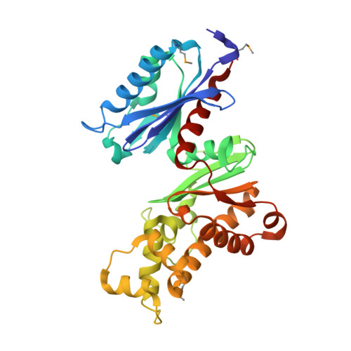 | |
UniProt | |||||
Entity Groups | |||||
| Sequence Clusters | 30% Identity50% Identity70% Identity90% Identity95% Identity100% Identity | ||||
| UniProt Group | Q830J4 | ||||
Sequence AnnotationsExpand | |||||
Reference Sequence | |||||
| Ligands 5 Unique | |||||
|---|---|---|---|---|---|
| ID | Chains | Name / Formula / InChI Key | 2D Diagram | 3D Interactions | |
| PEG Download:Ideal Coordinates CCD File | V [auth D] | DI(HYDROXYETHYL)ETHER C4 H10 O3 MTHSVFCYNBDYFN-UHFFFAOYSA-N |  | ||
| SO4 Download:Ideal Coordinates CCD File | T [auth D], U [auth D] | SULFATE ION O4 S QAOWNCQODCNURD-UHFFFAOYSA-L |  | ||
| GOL Download:Ideal Coordinates CCD File | G [auth A] H [auth A] I [auth A] L [auth B] P [auth C] | GLYCEROL C3 H8 O3 PEDCQBHIVMGVHV-UHFFFAOYSA-N |  | ||
| ZN Download:Ideal Coordinates CCD File | E [auth A], J [auth B], M [auth C], R [auth D] | ZINC ION Zn PTFCDOFLOPIGGS-UHFFFAOYSA-N |  | ||
| MG Download:Ideal Coordinates CCD File | F [auth A], K [auth B], N [auth C], O [auth C], S [auth D] | MAGNESIUM ION Mg JLVVSXFLKOJNIY-UHFFFAOYSA-N |  | ||
| Modified Residues 1 Unique | |||||
|---|---|---|---|---|---|
| ID | Chains | Type | Formula | 2D Diagram | Parent |
| MSE Query on MSE | A, B, C, D | L-PEPTIDE LINKING | C5 H11 N O2 Se |  | MET |
| Length ( Å ) | Angle ( ˚ ) |
|---|---|
| a = 62.083 | α = 90 |
| b = 131.26 | β = 92.48 |
| c = 82.763 | γ = 90 |
| Software Name | Purpose |
|---|---|
| REFMAC | refinement |
| SBC-Collect | data collection |
| HKL-3000 | data collection |
| HKL-2000 | data reduction |
| HKL-2000 | data scaling |
| autoSHARP | phasing |
| PHENIX | phasing |


