2QDT | pdb_00002qdt

Structural Basis for the Broad-Spectrum Inhibition of Metallo-{Beta}-Lactamases: L1- IS38 Complex


Experimental Data Snapshot

  • Method: X-RAY DIFFRACTION
  • Resolution: 2.00 Å
  • R-Value Free: 
    0.192 (Depositor), 0.202 (DCC) 
  • R-Value Work: 
    0.170 (Depositor), 0.180 (DCC) 
  • R-Value Observed: 
    0.171 (Depositor) 

Starting Model: experimental
View more details

wwPDB Validation 3D Report Full Report

Validation slider image for 2QDT

Ligand Structure Quality Assessment 


This is version 1.3 of the entry. See complete history

Literature

Structural basis for the broad-spectrum inhibition of metallo-beta-lactamases by thiols.

Lienard, B.M.Garau, G.Horsfall, L.Karsisiotis, A.I.Damblon, C.Lassaux, P.Papamicael, C.Roberts, G.C.Galleni, M.Dideberg, O.Frere, J.M.Schofield, C.J.

(2008) Org Biomol Chem 6: 2282-2294

  • DOI: https://doi.org/10.1039/b802311e
  • Primary Citation Related Structures: 
    2QDS, 2QDT

  • PubMed Abstract: 

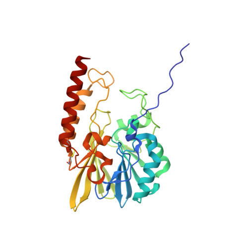
    The development of broad-spectrum metallo-beta-lactamase (MBL) inhibitors is challenging due to structural diversity and differences in metal utilisation by these enzymes. Analysis of structural data, followed by non-denturing mass spectrometric analyses, identified thiols proposed to inhibit representative MBLs from all three sub-classes: B1, B2 and B3. Solution analyses led to the identification of broad spectrum inhibitors, including potent inhibitors of the CphA MBL (Aeromonas hydrophila). Structural studies revealed that, as observed for other B1 and B3 MBLs, inhibition of the L1 MBL thiols involves metal chelation. Evidence is reported that this is not the case for inhibition of the CphA enzyme by some thiols; the crystal structure of the CphA-Zn-inhibitor complex reveals a binding mode in which the thiol does not interact with the zinc. The structural data enabled the design and the production of further more potent inhibitors. Overall the results suggest that the development of reasonably broad-spectrum MBL inhibitors should be possible.


  • Organizational Affiliation
    • Chemistry Research Laboratory and OCISB, University of Oxford, 12 Mansfield Road, Oxford, OX1 3TA, UK.

Macromolecule Content 

  • Total Structure Weight: 29.4 kDa 
  • Atom Count: 2,277 
  • Modeled Residue Count: 266 
  • Deposited Residue Count: 269 
  • Unique protein chains: 1

Macromolecules

Find similar proteins by:|  3D Structure
Entity ID: 1
MoleculeChains  Sequence LengthOrganismDetailsImage
Metallo-beta-lactamase L1269Stenotrophomonas maltophiliaMutation(s): 0 
Gene Names: L1
EC: 3.5.2.6
UniProt
Find proteins for P52700 (Stenotrophomonas maltophilia)
Explore P52700 
Go to UniProtKB:  P52700
Entity Groups
Sequence Clusters30% Identity50% Identity70% Identity90% Identity95% Identity100% Identity
UniProt GroupP52700
Sequence Annotations
Expand
Reference Sequence

Small Molecules

Ligands 3 Unique
IDChains Name / Formula / InChI Key2D Diagram3D Interactions
I38

Query on I38


Download:Ideal Coordinates CCD File
H [auth A]N-(3-MERCAPTOPROPANOYL)-D-ALANINE
C6 H11 N O3 S
JIAFOCJABIEPNM-SCSAIBSYSA-N
SO4

Query on SO4


Download:Ideal Coordinates CCD File
E [auth A],
F [auth A],
G [auth A]
SULFATE ION
O4 S
QAOWNCQODCNURD-UHFFFAOYSA-L
ZN

Query on ZN


Download:Ideal Coordinates CCD File
B [auth A],
C [auth A],
D [auth A]
ZINC ION
Zn
PTFCDOFLOPIGGS-UHFFFAOYSA-N

Experimental Data & Validation

Experimental Data

  • Method: X-RAY DIFFRACTION
  • Resolution: 2.00 Å
  • R-Value Free:  0.192 (Depositor), 0.202 (DCC) 
  • R-Value Work:  0.170 (Depositor), 0.180 (DCC) 
  • R-Value Observed: 0.171 (Depositor) 
Space Group: P 64 2 2
Unit Cell:
Length ( Å )Angle ( ˚ )
a = 105.319α = 90
b = 105.319β = 90
c = 97.96γ = 120
Software Package:
Software NamePurpose
CCP4model building
REFMACrefinement
MAR345dtbdata collection
MOSFLMdata reduction
SCALAdata scaling
CCP4phasing

Structure Validation

View Full Validation Report


Ligand Structure Quality Assessment 


Entry History 

Deposition Data

Revision History  (Full details and data files)

  • Version 1.0: 2007-08-28
    Type: Initial release
  • Version 1.1: 2011-07-13
    Changes: Derived calculations, Version format compliance
  • Version 1.2: 2023-08-30
    Changes: Data collection, Database references, Derived calculations, Refinement description
  • Version 1.3: 2024-10-30
    Changes: Structure summary