Structural, Functional, and Mutational Analysis of the NblA Protein Provides Insight into Possible Modes of Interaction with the Phycobilisome
Dines, M., Sendersky, E., David, L., Schwarz, R., Adir, N.(2008) J Biological Chem 283: 30330-30340
- PubMed: 18718907 Search on PubMedSearch on PubMed Central
- DOI: https://doi.org/10.1074/jbc.M804241200
- Primary Citation Related Structures:
2Q8V, 2QDO, 3CS5 - PubMed Abstract:
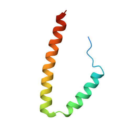
The enormous macromolecular phycobilisome antenna complex (>4 MDa) in cyanobacteria and red algae undergoes controlled degradation during certain forms of nutrient starvation. The NblA protein (approximately 6 kDa) has been identified as an essential component in this process. We have used structural, biochemical, and genetic methods to obtain molecular details on the mode of action of the NblA protein. We have determined the three-dimensional structure of the NblA protein from both the thermophilic cyanobacterium Thermosynechococcus vulcanus and the mesophilic cyanobacterium Synechococcus elongatus sp. PCC 7942. The NblA monomer has a helix-loop-helix motif which dimerizes into an open, four-helical bundle, identical to the previously determined NblA structure from Anabaena. Previous studies indicated that mutations to NblA residues near the C terminus impaired its binding to phycobilisome proteins in vitro, whereas the only mutation known to affect NblA function in vivo is located near the protein N terminus. We performed random mutagenesis of the S. elongatus nblA gene which enabled the identification of four additional amino acids crucial for NblA function in vivo. This data shows that essential amino acids are not confined to the protein termini. We also show that expression of the Anabaena nblA gene complements phycobilisome degradation in an S. elongatus NblA-null mutant despite the low homology between NblAs of these cyanobacteria. We propose that the NblA interacts with the phycobilisome via "structural mimicry" due to similarity in structural motifs found in all phycobiliproteins. This suggestion leads to a new model for the mode of NblA action which involves the entire NblA protein.
- Schulich Faculty of Chemistry and Institute of Catalysis, Science, and Technology, Technion-Israel Institute of Technology, Technion City, Haifa 32000, Israel.
Organizational Affiliation: