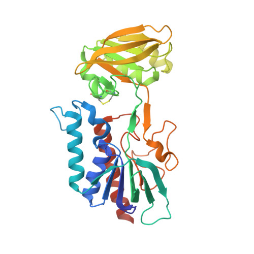
Thioredoxin system from Deinococcus radiodurans.
Obiero, J., Pittet, V., Bonderoff, S.A., Sanders, D.A.(2010) J Bacteriol 192: 494-501
- PubMed: 19933368
- DOI: https://doi.org/10.1128/JB.01046-09
- Primary Citation of Related Structures:
2Q7V - PubMed Abstract:
This paper describes the cloning, purification, and characterization of thioredoxin (Trx) and thioredoxin reductase (TrxR) and the structure determination of TrxR from the ionizing radiation-tolerant bacterium Deinococcus radiodurans strain R1. The genes from D. radiodurans encoding Trx and TrxR were amplified by PCR, inserted into a pET expression vector, and overexpressed in Escherichia coli. The overexpressed proteins were purified by metal affinity chromatography, and their activity was demonstrated using well-established assays of insulin precipitation (for Trx), 5,5'-dithiobis(2-nitrobenzoic acid) (DTNB) reduction, and insulin reduction (for TrxR). In addition, the crystal structure of oxidized TrxR was determined at 1.9-A resolution. The overall structure was found to be very similar to that of E. coli TrxR and homodimeric with both NADPH- and flavin adenine dinucleotide (FAD)-binding domains containing variants of the canonical nucleotide binding fold, the Rossmann fold. The K(m) (5.7 muM) of D. radiodurans TrxR for D. radiodurans Trx was determined and is about twofold higher than that of the E. coli thioredoxin system. However, D. radiodurans TrxR has a much lower affinity for E. coli Trx (K(m), 44.4 muM). Subtle differences in the surface charge and shape of the Trx binding site on TrxR may account for the differences in recognition. Because it has been suggested that TrxR from D. radiodurans may have dual cofactor specificity (can utilize both NADH and NADPH), D. radiodurans TrxR was tested for its ability to utilize NADH as well. Our results show that D. radiodurans TrxR can utilize only NADPH for activity.
- Department of Chemistry, University of Saskatchewan, Saskatoon S7N 5C9, Canada.
Organizational Affiliation:


