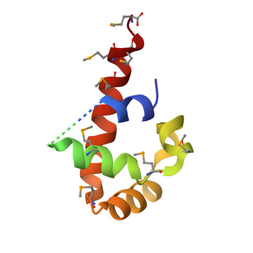
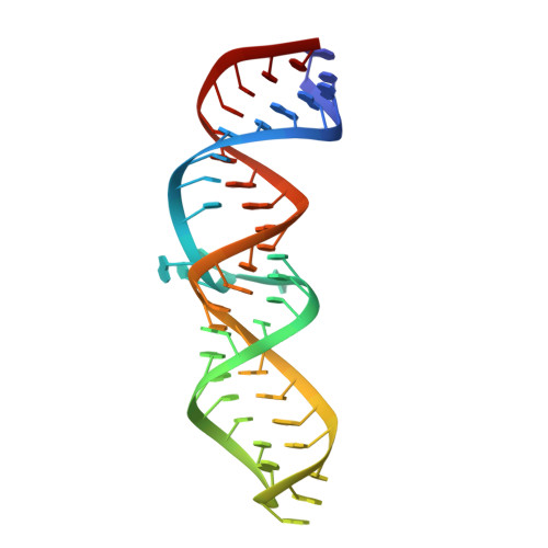
2PXL

Variant 9 of Ribonucleoprotein Core of the E. Coli Signal Recognition Particle


Experimental Data Snapshot

  • Method: X-RAY DIFFRACTION
  • Resolution: 2.50 Å
  • R-Value Free: 0.260 
  • R-Value Work: 0.237 

wwPDB Validation   3D Report Full Report


This is version 1.3 of the entry. See complete history


Literature

A General Strategy to Solve the Phase Problem in RNA Crystallography.

Keel, A.Y.Rambo, R.P.Batey, R.T.Kieft, J.S.

(2007) Structure 15: 761-772

  • DOI: https://doi.org/10.1016/j.str.2007.06.003
  • Primary Citation of Related Structures:  
    2PXB, 2PXD, 2PXE, 2PXF, 2PXK, 2PXL, 2PXP, 2PXQ, 2PXT, 2PXU, 2PXV

  • PubMed Abstract: 

    X-ray crystallography of biologically important RNA molecules has been hampered by technical challenges, including finding heavy-atom derivatives to obtain high-quality experimental phase information. Existing techniques have drawbacks, limiting the rate at which important new structures are solved. To address this, we have developed a reliable means to localize heavy atoms specifically to virtually any RNA. By solving the crystal structures of thirteen variants of the G*U wobble pair cation binding motif, we have identified a version that when inserted into an RNA helix introduces a high-occupancy cation binding site suitable for phasing. This "directed soaking" strategy can be integrated fully into existing RNA crystallography methods, potentially increasing the rate at which important structures are solved and facilitating routine solving of structures using Cu-Kalpha radiation. This method already has been used to solve several crystal structures.


  • Organizational Affiliation

    Department of Biochemistry and Molecular Genetics, University of Colorado at Denver and Health Sciences Center, Aurora, CO 80045, USA.


Macromolecules

Find similar proteins by:  (by identity cutoff)  |  3D Structure
Entity ID: 2
MoleculeChains Sequence LengthOrganismDetailsImage
Signal recognition particle proteinB [auth A]102Escherichia coliMutation(s): 14 
Gene Names: ffh
EC: 3.6.5.4
UniProt
Find proteins for P0AGD7 (Escherichia coli (strain K12))
Explore P0AGD7 
Go to UniProtKB:  P0AGD7
Entity Groups  
Sequence Clusters30% Identity50% Identity70% Identity90% Identity95% Identity100% Identity
UniProt GroupP0AGD7
Sequence Annotations
Expand
  • Reference Sequence
Find similar nucleic acids by:  (by identity cutoff)  |  3D Structure
Entity ID: 1
MoleculeChains LengthOrganismImage
4.5 S RNAA [auth B]47N/A
Sequence Annotations
Expand
  • Reference Sequence
Experimental Data & Validation

Experimental Data

  • Method: X-RAY DIFFRACTION
  • Resolution: 2.50 Å
  • R-Value Free: 0.260 
  • R-Value Work: 0.237 
  • Space Group: C 1 2 1
Unit Cell:
Length ( Å )Angle ( ˚ )
a = 122.688α = 90
b = 78.847β = 95.91
c = 32.46γ = 90
Software Package:
Software NamePurpose
d*TREKdata scaling
CNSrefinement
PDB_EXTRACTdata extraction
CrystalCleardata collection
d*TREKdata reduction
CNSphasing

Structure Validation

View Full Validation Report


Entry History 

Deposition Data

Revision History  (Full details and data files)

  • Version 1.0: 2007-08-07
    Type: Initial release
  • Version 1.1: 2011-07-13
    Changes: Version format compliance
  • Version 1.2: 2018-03-07
    Changes: Data collection
  • Version 1.3: 2021-10-20
    Changes: Database references, Derived calculations