2PLT

STRUCTURE DETERMINATION OF PLASTOCYANIN FROM A CRYSTAL SPECIMEN WITH HEMIHEDRAL TWINNING FRACTION OF ONE-HALF


Experimental Data Snapshot

  • Method: X-RAY DIFFRACTION
  • Resolution: 1.50 Å
  • R-Value Work: 0.168 
  • R-Value Observed: 0.168 

wwPDB Validation   3D Report Full Report


This is version 1.4 of the entry. See complete history


Literature

The 1.5-A crystal structure of plastocyanin from the green alga Chlamydomonas reinhardtii.

Redinbo, M.R.Cascio, D.Choukair, M.K.Rice, D.Merchant, S.Yeates, T.O.

(1993) Biochemistry 32: 10560-10567

  • DOI: https://doi.org/10.1021/bi00091a005
  • Primary Citation of Related Structures:  
    2PLT

  • PubMed Abstract: 

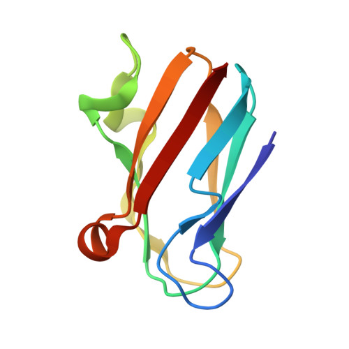
    The crystal structure of plastocyanin from the green alga Chlamydomonas reinhardtii has been determined at 1.5-A resolution with a crystallographic R factor of 16.8%. Plastocyanin is a small (98 amino acids), blue copper-binding protein that catalyzes the transfer of electrons in oxygenic photosynthesis from cytochrome f in the quinol oxidase complex to P700+ in photosystem I. Chlamydomonas reinhardtii plastocyanin is an eight-stranded, antiparallel beta-barrel with a single copper atom coordinated in quasitetrahedral geometry by two imidazole nitrogens (from His-37 and His-87), a cysteine sulfur (from Cys-84), and a methionine sulfur (from Met-92). The molecule contains a region of negative charge surrounding Tyr-83 (the putative distant site of electron transfer) and an exclusively hydrophobic region surrounding His-87; these regions are thought to be involved in the recognition of reaction partners for the purpose of directing electron transfer. Chlamydomonas reinhardtii plastocyanin is similar to the other plastocyanins of known structure, particularly the green algal plastocyanins from Enteromorpha prolifera and Scenedesmus obliquus. A potential "through-bond" path of electron transfer has been identified in the protein that involves the side chain of Tyr-83, the main-chain atoms between residues 83 and 84, the side chain of Cys-84, the copper atom, and the side chain of His-87.


  • Organizational Affiliation

    Department of Chemistry and Biochemistry and Molecular Biology Institute, University of California at Los Angeles, California 90024.


Macromolecules
Find similar proteins by:  (by identity cutoff)  |  3D Structure
Entity ID: 1
MoleculeChains Sequence LengthOrganismDetailsImage
PLASTOCYANIN98Chlamydomonas reinhardtiiMutation(s): 0 
Membrane Entity: Yes 
UniProt
Find proteins for P18068 (Chlamydomonas reinhardtii)
Explore P18068 
Go to UniProtKB:  P18068
Entity Groups  
Sequence Clusters30% Identity50% Identity70% Identity90% Identity95% Identity100% Identity
UniProt GroupP18068
Sequence Annotations
Expand
  • Reference Sequence
Small Molecules
Ligands 2 Unique
IDChains Name / Formula / InChI Key2D Diagram3D Interactions
CU
Query on CU

Download Ideal Coordinates CCD File 
B [auth A]COPPER (II) ION
Cu
JPVYNHNXODAKFH-UHFFFAOYSA-N
CA
Query on CA

Download Ideal Coordinates CCD File 
C [auth A]CALCIUM ION
Ca
BHPQYMZQTOCNFJ-UHFFFAOYSA-N
Experimental Data & Validation

Experimental Data

  • Method: X-RAY DIFFRACTION
  • Resolution: 1.50 Å
  • R-Value Work: 0.168 
  • R-Value Observed: 0.168 
  • Space Group: P 32
Unit Cell:
Length ( Å )Angle ( ˚ )
a = 61.8α = 90
b = 61.8β = 90
c = 25.2γ = 120
Software Package:
Software NamePurpose
X-PLORmodel building
X-PLORrefinement
X-PLORphasing

Structure Validation

View Full Validation Report


Entry History 

Deposition Data

Revision History  (Full details and data files)

  • Version 1.0: 1993-10-31
    Type: Initial release
  • Version 1.1: 2008-03-24
    Changes: Version format compliance
  • Version 1.2: 2011-07-13
    Changes: Version format compliance
  • Version 1.3: 2012-07-18
    Changes: Database references
  • Version 1.4: 2024-02-21
    Changes: Data collection, Database references, Derived calculations, Other