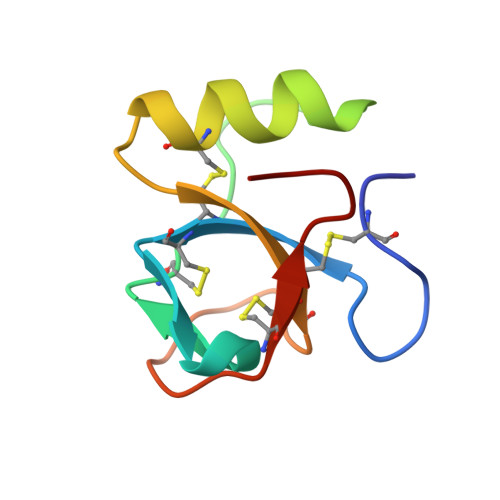
2PL6

Monoclinic crystal structure of hydrophobin HFBII in presence of a detergent


Experimental Data Snapshot

  • Method: X-RAY DIFFRACTION
  • Resolution: 2.20 Å
  • R-Value Free: 0.280 
  • R-Value Work: 0.224 

Starting Model: experimental
View more details

wwPDB Validation   3D Report Full Report


Ligand Structure Quality Assessment 


This is version 1.5 of the entry. See complete history


Literature

Crystal Structures of Hydrophobin HFBII in the Presence of Detergent Implicate the Formation of Fibrils and Monolayer Films.

Kallio, J.M.Linder, M.B.Rouvinen, J.

(2007) J Biol Chem 282: 28733-28739

  • DOI: https://doi.org/10.1074/jbc.M704238200
  • Primary Citation of Related Structures:  
    2PL6, 2PL7

  • PubMed Abstract: 

    Hydrophobins are small, amphiphilic proteins secreted by filamentous fungi. Their functionality arises from a patch of hydrophobic residues on the protein surface. Spontaneous self-assembly of hydrophobins leads to the formation of an amphiphilic layer that remarkably reduces the surface tension of water. We have determined by x-ray diffraction two new crystal structures of Trichoderma reesei hydrophobin HFBII in the presence of a detergent. The monoclinic crystal structure (2.2A resolution, R = 22, R(free) = 28) is composed of layers of hydrophobin molecules where the hydrophobic surface areas of the molecules are aligned within the layer. Viewed perpendicular to the aligned hydrophobic surface areas, the molecules in the layer pack together to form six-membered rings, thus leaving small pores in the layer. Similar packing has been observed in the atomic force microscopy images of the self-assembled layers of class II hydrophobin, indicating that the crystal structure resembles that of natural hydrophobin film. The orthorhombic crystal structure (1.0 A resolution, R = 13, R(free) = 15) is composed of fiber-like arrays of protein molecules. Rodlet structures have been observed on amphiphilic layers formed by class I hydrophobins; fibrils of class II hydrophobins appear by vigorous shaking. We propose that the structure of the fibrils and/or rodlets is similar to that observed in the crystal structure.


  • Organizational Affiliation

    Department of Chemistry, University of Joensuu, P. O. Box 111, 80101 Joensuu, Finland. Electronic address: johanna.kallio@joensuu.fi.


Macromolecules
Find similar proteins by:  (by identity cutoff)  |  3D Structure
Entity ID: 1
MoleculeChains Sequence LengthOrganismDetailsImage
Hydrophobin-2
A, B, C, D, E
A, B, C, D, E, F, G, H
71Trichoderma reeseiMutation(s): 0 
UniProt
Find proteins for P79073 (Hypocrea jecorina)
Explore P79073 
Go to UniProtKB:  P79073
Entity Groups  
Sequence Clusters30% Identity50% Identity70% Identity90% Identity95% Identity100% Identity
UniProt GroupP79073
Sequence Annotations
Expand
  • Reference Sequence
Experimental Data & Validation

Experimental Data

  • Method: X-RAY DIFFRACTION
  • Resolution: 2.20 Å
  • R-Value Free: 0.280 
  • R-Value Work: 0.224 
  • Space Group: P 1 21 1
Unit Cell:
Length ( Å )Angle ( ˚ )
a = 61.08α = 90
b = 66.41β = 99.23
c = 79.77γ = 90
Software Package:
Software NamePurpose
XSCALEdata scaling
PHASERphasing
CNSrefinement
PDB_EXTRACTdata extraction
ADSCdata collection
XDSdata reduction

Structure Validation

View Full Validation Report


Ligand Structure Quality Assessment 


Entry History 

Deposition Data

Revision History  (Full details and data files)

  • Version 1.0: 2007-07-17
    Type: Initial release
  • Version 1.1: 2007-09-27
    Changes: Version format compliance
  • Version 1.2: 2011-07-13
    Changes: Version format compliance
  • Version 1.3: 2020-07-29
    Type: Remediation
    Reason: Carbohydrate remediation
    Changes: Derived calculations, Structure summary
  • Version 1.4: 2023-08-30
    Changes: Data collection, Database references, Refinement description, Structure summary
  • Version 1.5: 2024-10-30
    Changes: Structure summary